CELL MEMBRANES-STRUCTURE AND TRANSPORT
THE FLUID MOSAIC MODEL
Introduction
One of the challenges faced by all living things, be they amoebae or humans, is to separate their internal environment from the external environment. Critical nutrients must get into the cells and wastes must get out. To make matters more complex, cells need to be able to regulate that movement, letting the materials cross at some times and preventing them from crossing at others. Another challenge is finding a way for cells to communicate with each other. Cells in the brain, for example, need to be able to tell cells in the heart to beat faster. The solution to these challenges lies in the properties of the cell membrane (plasmamembrane). This delicate structure is essential to the life of cells. When the membrane loses its ability to carry out these processes, the cell dies.
In this lesson we will study this amazing structure. We will learn how it allows some things to readily cross and prevents others. Hopefully you will gain an appreciation of its complexity and come to realize how important it is to cellular function.
Fluid Mosaic Model of the Membrane
The plasma membrane is more than just a sack to hold the contents of the cell. It plays an important role in cellular function and the maintenance of homeostasis. One obvious function is to regulate what enters and leaves the cell. This process is highly coordinated and very specific. In addition, the cell membrane responds to countless chemical messengers in ways that alter the activity of the cell. As we discuss the structure of the plasma membrane, keep in mind that this description also applies to other membranes that are components of intracellular organelles.
Our modern model of the cell membrane is called the "Fluid Mosaic Model of the Cell Membrane." The word, fluid implies that the membrane is constantly changing and moving. Indeed, it is not a static structure, but one that changes as the cellular needs change. A good example of this fluidity can be seen with the uptake of glucose into muscle cells. The plasma membrane of these cells is not normally permeable to glucose, preventing it from entering the cell. Only when a signal molecule, insulin, is present can glucose enter. The presence of this signal results in the insertion of special glucose transporters into the membrane, allowing glucose to enter the cell. When insulin is no longer there, the carriers are removed, demonstrating the ability of the membrane to change, depending on the needs of the cell. Additionally, components of the membrane are not rigidly fixed in one area but often have the freedom to move laterally within the membrane. The term, mosaic conjures up an image of a picture composed of numerous small, and different pieces. Indeed, the membrane contains many different components that make up the intact membrane. The following link shows the structure and function of the membrane: http://www.youtube.com/watch?v=Qqsf_UJcfBc(Transcription Available).
COMPOSITION OF THE MEMBRANE
Lipids
A key component of the membrane is a double layer of phospholipids, the phospholipid bilayer. This bilayer forms the scaffolding into which the other components of the membrane are housed. Recall phospholipids are composed of a hydrophilic head containing a phosphate group and two hydrophobic tails composed of long chain fatty acids. This bilayer has a central hydrophobic region and two outer hydrophilic sections, one facing the aqueous interior of the cell, and one facing the aqueous extracellular space (see figure below).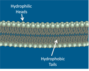
Image created by BYU-IU student, Hannah Crowder 2013
In water, phospholipids can form a bilayer. The hydrophobic fatty acid tails turn away from the water and the hydrophilic phosphatesheads turn towards the water.
The hydrophobic core of the membrane creates a barrier, preventing the movement of hydrophilic substance such as ions and large polar molecules across the membrane. Hydrophobic (lipid soluble) materials, on the other hand typically move readily across the membrane. Because some things easily pass through the membrane and others do not, we describe the membrane as being selectively permeable. The following link may help you better understand the concept of selective permeability:
http://www.youtube.com/watch?v=5jZXCDcM14g(Transcription Available)
In addition to the phospholipids, another important lipid found in membranes is cholesterol. Cholesterol is a hydrophobic molecule and resides among the fatty acids tails of the phospholipid bilayer. As mentioned above, the membrane exhibits fluidity allowing movement of components within the membrane. Cholesterol plays an important role in helping regulate the fluidity of the membrane by limiting lateral movement of the phospholipids. This is very important because if the membrane loses fluidity or becomes too fluid, cellular function may be impaired. The presence of cholesterol allows the membrane to maintain proper fluidity over the range of temperatures our cells are exposed to. While it is true that our core body temperature remains fairly constant, temperatures in our extremities may vary considerably. Think of the range of temperatures the cells in your hands are exposed to. Recent evidence suggests that cholesterol may also play a role in cell signaling, due to its influence on the formation of specialized structures in the membranes, called lipid rafts. Together, phospholipids and cholesterol make nearly 50% of the membrane.
Proteins
Making up about another 50% of the membrane are the membrane proteins. The figure below demonstrates the relationship of the membrane proteins (blue) with the phospholipid bilayer (red). Note that some of the proteins are found only on the surface of the membrane. These are called peripheral or extrinsic proteins because they do not extend through the membrane. One function of the peripheral proteins is to attach the membrane to the cytoskeletal proteins inside the cell, or to proteins of the extracellular matrix. For example, the cells lining the blood vessels utilize peripheral proteins to attach to the tissues outside the vessel, thus, holding the cells in place.As seen in the figure below, other proteins pass all the way through the membrane.
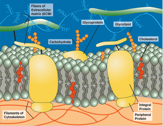
Image created by BYU-IU student, Hannah Crowder 2013
Model of the cell membrane demonstrating the relationships between the lipids, proteins, and carbohydrates.
These proteins are called integral or intrinsic proteins and have segments that associate with the hydrophobic region of the membrane. These integral proteins perform a number of important functions in the cell. Based on their functions, these integral proteins can be grouped into the following categories:
Transport proteins
Integral proteins can act as channels allowing hydrophilic materials, such as ions, to cross the membrane. You can picture these proteins as looking like tubes between the interior and exterior of the cell (see figure below). These channel proteins are usually gated, meaning that, like a door, they allow substances to cross only when they are open. We will have more to say about channel gating later.
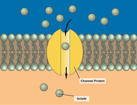
Image created by BYU-IU student, Hannah Crowder 2013
Channel proteins allow solutes, such as ions, to move across the membrane.
Another type of transport proteins are carrier proteins (see next figure below). Carriers have sites that bind to specific solutes. For example, one type of carrier binds with glucose, while another carrier binds to urea. Once the solute binds, the carrier protein changes shape, allowing the solute to move across the membrane. Imagine a revolving door. As these doors turn, they are open to either the inside of the building or to the outside, but never to both at the same time. You can enter a revolving door from the outside of a room and move the door so that it is now open to the inside of the room. At no time, in this process was the door open to both sides at the same time. This is, essentially, how carrier proteins work. Rather than pushing against the door, the binding of the solute causes the protein to change shape and open to the opposite side of the membrane. Carrier proteins bind to solutes and then move them across the membrane by changing shape.
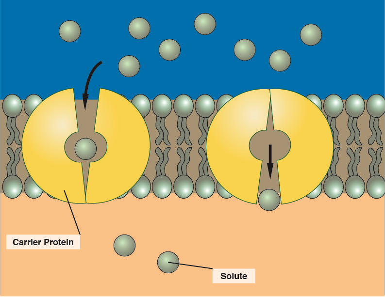
Image created by BYU-IU student, Hannah Crowder 2013
Enzymes
Integral membrane proteins can function as enzymes, catalyzing important chemical reactions. The enzyme, lactase that digests the disaccharide lactose in the small intestine, is an integral membrane protein in the cells that line the lumen of the duodenum. Lack of, or insufficient amounts of this enzyme is responsible for the discomfort associated with lactose intolerance.
Receptor Proteins
Integral proteins may act as receptors and allow the cell to respond to chemical messengers which regulate the activities of the cell. For example, epinephrine (adrenaline) binds to receptors on cardiac muscle cells causing your heart to beat faster when you are frightened.
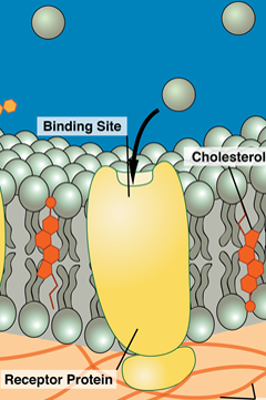
Image created by BYU-IU student, Hannah Crowder 2013
An important function of membrane proteins is to act as receptors that respond to chemical signals. When a chemical signal binds to one of these receptors, it transmits a signal to the inside of the cell that regulates cell function. As a very important example (that will come up multiple times in this course), we will look at one of the most abundant of these receptors, the G protein-coupled receptor (GPCR). The GPCR receptor complex is composed of two units: a receptor protein that binds to the chemical signal (the ligand), and the G-protein complex associated with the inner leaflet of the membrane. The receptor passes all the way through the membrane with a ligand binding site on the external surface and a G protein binding site on the internal surface. The G protein complex is composed of three subunits called the alpha, beta, and gamma subunits. The alpha subunit has a site that can bind Guanosine Triphosphate (GTP) or Guanosine Diphosphate (GDP), hence the name "G" protein. At rest, the G protein is bound to GDP and the three subunits (alpha, beta, and gamma) are bound together. When a ligand binds to the receptor on the surface of the cell, the G protein binding site changes shape, allowing the G protein to bind to the receptor. This binding causes the G protein to change shape and the GDP exits the binding site on the alpha subunit and is replaced by GTP. The binding of GTP causes the alpha subunit to separate from the other two subunits (beta/gamma dimer). Once separated, the alpha subunit is activated and can turn on other processes inside the cell. The mechanism of action is typically mediated by one of two enzymes, adenylyl cyclase and phospholipase C. These signals initiate a cascade of events resulting in changes of cell function. Cellular responses include: activation of metabolic enzymes, opening or closing ion channels, turning on transporters, initiating gene transcription, regulating motility, regulating contractility, stimulating secretion, even controlling memory. After a short period of time, the GTP is broken down to GDP and phosphate allowing the alpha subunit to reunite with the beta/gamma dimer, turning off the signal.
To date, approximately 800 genes for G protein-coupled receptors have been identified. G-proteins are very common in physiology and it is important to study the details known about this receptor. Ligand activation and the detailed mechanism of effect on the G-protein is illustrated in the following figure.
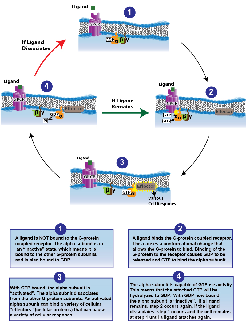
Image drawn by JS Falll 2014
Attachment Proteins
Integral proteins are involved in attaching cells to each other as well as to the extracellular matrix and to intracellular structural proteins. Often a peripheral protein functions as a link between the integral proteins and the structural proteins or the matrix. These attachments give cells their strength and shape. The inability to form these connections can result in a number of pathological conditions, including muscular dystrophy.
These proteins allow cells to identify one another. Functions of these marker proteins include the ability of sperm cells to recognize the oocyte during fertilization, as well as the ability of our immune cells to distinguish between our own cells and a foreign cell, such as a bacterial cell, that might be trying to invade our bodies.
Carbohydrates
In addition to lipids and proteins, the membranes also contain carbohydrates. These are short-chained polysaccharides (oligosaccharides) that attach to the proteins and lipids on the extracellular layer of the membrane. If attached to a protein, they are called glycoproteins, and if attached to a lipid, they are called glycolipids. One function of the carbohydrates is to participate with the proteins in forming specific markers. Your blood type (A, B, AB or O), for example, is determined by the carbohydrates attached to proteins on your red blood cells. Additionally, some cells, such as the apical surface of epithelial cells have a dense layer of glycoprotein referred to as the glycocalyx. The glycocalyx has been implicated in cell recognition during development, adherence of cells to each other, and playing a role in the permeability of the membranes.
**You may use the buttons below to go to the next reading in this Module**

