Special Senses
THE SENSE OF VISION
The Nature of Light
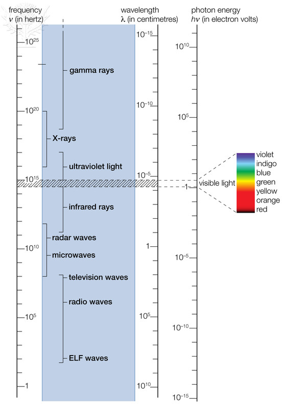
The function of the eye is to convert light waves to action potentials. In order to understand how this happens, we need to know a little about the nature of light. Visible light is a very small portion of the spectrum of electromagnetic radiation. The entire spectrum of electromagnetic radiation is shown in the image below.

© 2013 Encyclopædia Britannica, Inc. Downloaded from Image Quest Britannica; BYU-Idaho.
Spectrum of electromagnetic radiation. The visible spectrum is shown as colors.

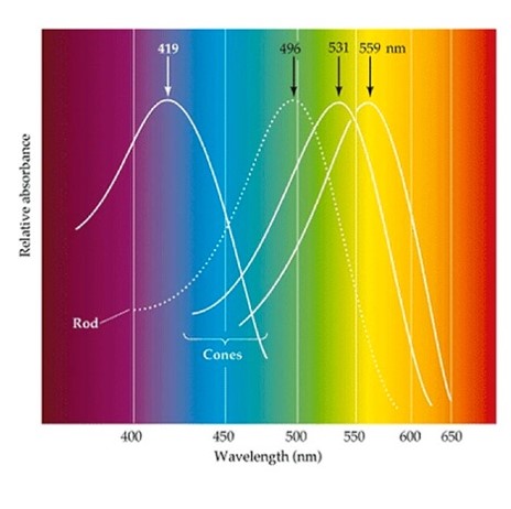
Title: Spectrum.jpeg; Author: http://webvision.med.utah.edu/; Site: http://webvision.med.utah.edu/book/part-ii-anatomy-and-physiology-of-the-retina/photoreceptors/; License: Copyright © 2015 Webvision: Attribution, Noncommercial, No Derivative Works Creative Commons license.
The image above shows the peak absorption of each of the cone cells as well as the rod cells.
The nature of electromagnetic radiation, and hence visible light, cannot be described using a single model. Some of light’s properties can be explained by describing it as a wave. For example the color of light that we perceive is based on the wavelength of the light waves. However, other properties suggest that light exists as discrete packets of energy called photons. The image above shows the relationship between wavelength and the energy in a photon of light. The shorter the wavelength, the greater the energy. Hence, gamma waves have very short wavelengths and contain large amounts of energy while radio waves have very long wavelengths but relatively small amounts of energy. The portion of the spectrum of electromagnetic radiation that we can perceive is referred to as the visible spectrum and includes light with wavelengths between 380 (violet) and 700 nm (red).
When light strikes an object, one of three things will happen. If the object is transparent the light is transmitted, meaning it will pass through the object. However, if the object is not transparent the light will either be absorbed or it will be reflected. The color that we perceive as we look at an object is due to the light that is being reflected off of it. Hence, if we see yellow, the yellow wavelength light is being reflected and the other wavelengths are being absorbed. Objects that appear black to our eyes absorb all of the light that is striking them while objects that appear white reflect all of the light that is striking them.
The Retina
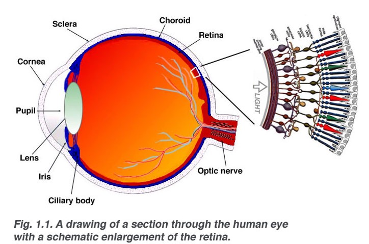
The structure of the eye responsible for converting light waves into action potentials is the retina. The neural layer of the retina is composed of three main types of cells: the photoreceptors, the bipolar neurons and the ganglion cells. The photoreceptors, as the name implies, have the responsibility of capturing the light and converting it to an electrical signal. There are two types of photoreceptors in the retina, the rods and the cones. The rods see only in black and white and are mainly responsible for our night vision. The cones, on the other hand, can see in color and are responsible for color vision as well as sharp vision. Each eye contains about 120,000,000 rods and 6,000,000 cones. Although they detect light of different wavelengths, structurally, rods and cones are similar. They are composed of an outer segment that touches the pigment epithelium and is composed of numerous flattened discs stacked on each other (think of a stack of dinner plates). The only difference is that in the rods, all of the discs are the same size while in the cones they gradually decrease in diameter as they move to the end of the cell. This results in the shape for which the cones were named. The outer segment connects to the inner segment which houses the nucleus and other organelles of the cell. The inner segment, in turn connects to the synaptic terminal which forms the connection between the photoreceptor and the bipolar neuron (see the images below).

Title: Sagschem.jpeg; Author: http://webvision.med.utah.edu/; Site: http://webvision.med.utah.edu/book/part-i-foundations/simple-anatomy-of-the-retina/; License: Copyright © 2015 Webvision: Attribution, Noncommercial, No Derivative Works Creative Commons license.

© 2013 Encyclopædia Britannica, Inc. Downloaded from Image Quest Britannica; BYU-Idaho.
Illustration of the photoreceptors of the eye, rods (black) and cones (green) and associated neurons within the neural retina. The brown cells are the bipolar neurons and the large orange structures are the cell bodies of the ganglion cells. The red colored layer at the top represents the choroid and the top purple layer the sclera.
The bipolar neurons, so named because they have one dendrite and one axon, are the connections between the photoreceptors and the ganglion cells. The axons of the ganglion cells form the optic nerve which exits the eye via the optic nerve. Two other cell types are shown in the image above. The horizontal cells can be seen in the layer where the photoreceptors synapse with the bipolar neurons and the Amacrine cells can be seen in the layer where the bipolar cells synapse with the ganglion cells. These two cells are involved in modulating the visual signals.
The distribution of the photoreceptors in the retina is not uniform. In the fovea centralis we find only cones. Moving move out from the fovea we start to see rods intermixed with the cones. The further out from the fovea we move the greater the number of rods and the fewer the number of cones.
In the image above note that the photoreceptors are located at the back of the retina. Light entering the eye must pass through the ganglion cells and the bipolar neurons before it gets to the photoreceptors. This doesn’t seem to be the best arrangement. However, at the fovea, the ganglion cells and the bipolar neurons radiate away from the cones in the fovea. Think of the crown of your head. All of the hairs radiate out from this point exposing the scalp. Because of this arrangement light striking the fovea has direct access to the photoreceptors, enhancing vision in this region of the retina.
Now lets examine the unique characteristics of the different photoreceptors, starting with the rods. The rods are very sensitive to light and will respond to a single photon of light. In addition they are part of convergent circuits in which several rods will converge on a single bipolar neuron and several bipolar neurons will converge on one ganglion cell. This allows for the summation of signals from several rods resulting in an action potential being sent to the brain. These properties make the rods ideal for seeing in very dim light, therefore rods are responsible for our night vision. During the day when there is plenty of light the rods are essentially inactivated due to a process called bleaching (more on this later). You are aware of how hard it is to see in a darkened theater when you first enter from bright light. After you have been there for a while and the rods become active we can see quite well in the room. Indeed, after about 40 minutes in the dark room our eyes are about 25,000 times more sensitive than they were when we first entered the room. There are two downsides to the use of rods, however. First, they do not see in color, rods see only in black and white. Second, due to the convergence their visual fields are quite large. Light striking any of the rods that converge on one ganglion cell will produce the same “pixel.” Therefore, vision with rods is very sensitive but not very acute (sharp). Cones, on the other hand, have essentially the opposite characteristics. First, there is very little convergence in their circuitry. Light striking two cones located next to each other would produce two different pixels in the brain. This allows for very sharp (acute) vision for images striking the fovea since there are only cones in the fovea. Second, the cones are much less sensitive than rods. At night, the intensity of light usually is not sufficient to stimulate the cones. Third, the cones are responsible for our color vision. We have three types of cones that respond to light in the red, green or blue wavelengths. By mixing the input from these three cones, humans can perceive about 1,000,000 different hues of color. You may know someone who is “color blind.” Color blindness is most often due to a genetic condition where the subject does not produce one or more of the cones. The most common condition is red-green colorblindness, where the person lacks either the red or green cone. Individuals with this condition can see colors, but they have a difficult time distinguishing between shades of green and red. The genes for the green and red cones are found on the X chromosome, therefore males have a much higher incidence of colorblindness since males only have one X chromosome. Women have two X chromosomes so even if they inherit a defective gene there is a good change the gene on the other X chromosome will be normal. For a women to be red-green colorblind, both her father and her mother would have to have the condition. Another type of colorblindness called Blue-yellow color blindness involves genes for the blue cones, but these genes are not on the X chromosome so it occurs at the same rate in both males and females.
Phototransduction
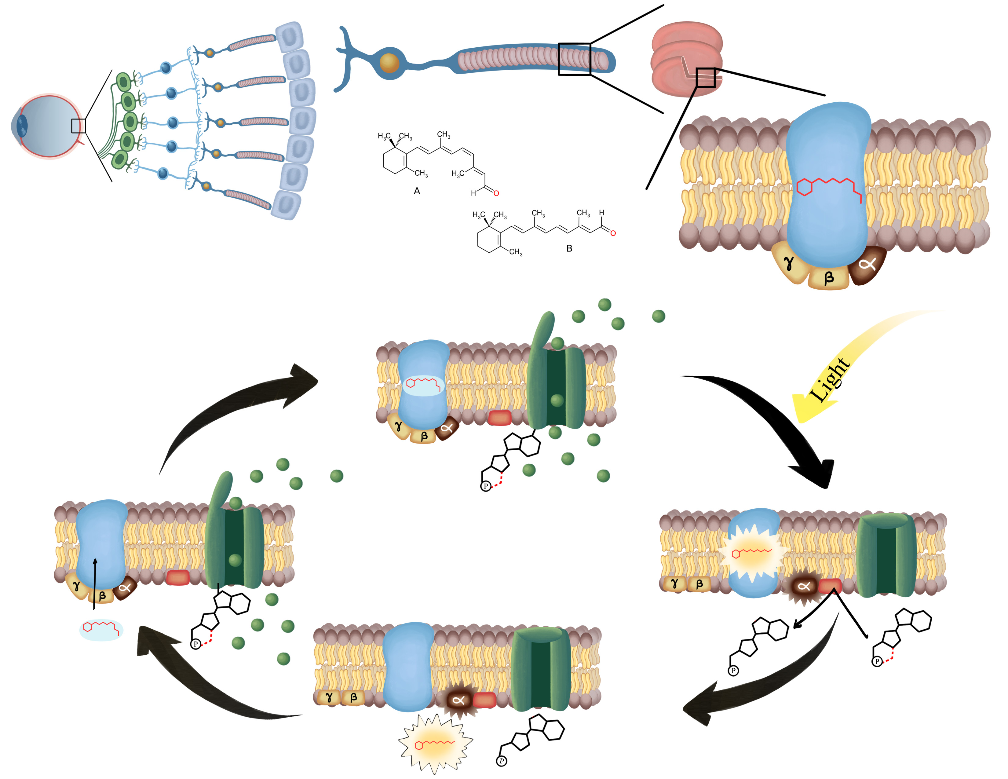
Now for the underlying question, how do the proteins that absorb photons of light produce the action potentials that travel to the brain to produce what we perceive as vision? This process is called phototransduction (see figure below). Since the process is essentially the same in both the rods and the cones we will look at the rods and then explain the subtle differences that occur in the cones. It all starts with the visual pigments that are embedded in the membranes of the disks found in the outer segment of the rods. This visual pigment is called rhodopsin and is composed of protein called opsin and a derivative of vitamin A called retinal. In the unexcited state, retinal has a bend in its hydrocarbon chain (11-cis retinal) and fits nicely in a binding site on the opsin. When light of the proper wavelength is absorbed by the visual pigment the energy of the light causes retinal to change shape and the hydrocarbon chain loses its bend (all-trans retinal) and no longer fits in the binding site. It should be noted that even though rods provide only non-color vision, light of the green wavelength is the most efficient in activating rhodopsin. When the retinal detaches from the opsin it becomes inactive. This process is known as bleaching. Opsin is actually a G-Protein coupled receptor (GPCR) that is activated by light, hence it is a photoreceptor. Once activated the GPCR activates the G-protein, separating the alpha subunit from the beta/gamma subunit (see module 5 for a review of GPCRs). In the photoreceptors of the eye the G-protein is called transducin. The alpha subunit then brings about a change in the cell. More on this later.

Created by BYU-I student Hannah Crowder, 2013
Phototransduction. The top image represents the photoreceptor in the dark. The green channel is the cGMP gated cation channel which is open and allowing cations (Na+ and Ca++) to depolarize the cell. When light strikes and changes the retinal from 11-cis to all-trans retinal it activates the G-protein transducin which results in the breakdown of cGMP and the closing of the cation channel. The cell will then hyperpolarize. Finally, All-trans retinal is converted back to 11-cis retinal and it re-attaches to opsin allowing cGPM to open the cation channel and once again depolarize the cell.
Photoreceptors are different than any receptors we have discussed to date in that they release neurotransmitter when they are not being stimulated. Here is how this works. There are three important important ion channels in the membranes of the photoreceptor cells, K+ leak channels, voltage-gated Ca2+ channels and cyclic GMP (cGMP) gated cation channels (Na+ and some Ca2+ move through this channel). When the photoreceptor is not being stimulated (in the dark), cGMP is bound to the cation channel and Na+ and Ca2+ diffuse into the cell maintaining it in a depolarized state. This depolarization causes the voltage gated Ca2+ channels to open, allowing more Ca2+ to diffuse into the cell. This Ca2+ triggers the release of the neurotransmitter glutamate by the process of exocytosis. The binding of glutamate to receptors on the bipolar neurons may be excitatory or inhibitory; it depends on what receptors are expressed on the bipolar neuron. In this module, we will focus on just the bipolar neurons that express receptors that cause inhibition when glutamate is attached.
When light is absorbed by rhodopsin and the G-protein is activated, the alpha subunit of the G-protein activates the enzyme phosphodiesterase. This enzyme breaks down cGMP to GMP. Once the cGMP is removed the cGMP-gated cation channels close and the membrane hyperpolarizes. This results in closing of the voltage-gated Ca2+ channels and glutamate release ceases. Removal of the inhibitory signal to the bipolar neurons allows them to fire and an action potential is sent to the brain. Eventually the G-protein is inactivated and phosphodiesterase is turned off. However, the rhodopsin cannot respond to light again until the retinal is returned to its bent, 11-cis, state. To do this, it diffuses into the pigment epithelium where enzymes act to restore the 11-cis state. It can then diffuse back into the rod cell and bind to opsin. The rod cell is ready to be activated again. The original bleaching process is very fast, fractions of seconds, but restoring the rhodopsin to its intact state can take several minutes. During the day, when we are exposed to sufficient light, the rhodopsin remains in the bleached state and the rods are essentially unresponsive to light. The mechanism is similar in the cones. The main difference is in the proteins of the visual pigment. The visual pigments in cones are similar to rhodopsin but they respond to different wavelengths of light allowing us to perceive different colors. Another difference, as stated above, is that the cones are much less sensitive to light. This is why the cones do well in full daylight when everything is brightly luminated. Finally, cones do not stay deactivated (bleached) as long as rods. Cones appear to be fairly resistant to large scale “bleaching” as they are able to recover 11-cis-retinal much more quickly so that at any given time there are at least some visual pigments ready for stimulation.
Recall that the axons of the ganglion cells form the optic nerves. These nerves enter the brain through the optic canals of the skull. As they move posteriorly they converge at a point just above the hypothalamus called the optic chiasm (the word chiasm comes from the Greek letter Chi or X, implying a point of crossing over). In the optic chiasm some of the axons cross to the opposite side of the brain while some stay on the same side. Let’s see if we can make sense of this. If we use the fovea as a reference point we can divide the retina into two halves, a lateral or temporal half and a medial or nasal half. Axons originating on the lateral retina enter the optic chiasm but do not cross over while those from the medial retina cross over to the opposite side of the brain. What are the implications of this? Suppose you are looking straight ahead and light from an image at your right enters your eye. It will be focused on the lateral retina of your left eye and the medial retina of your right eye. Since axons from the lateral retina do not cross over while axons from the medial retina do cross over, the image will be projected to only the left hemisphere of your occipital lobe. An object to your left would be projected to only your right hemisphere and the object directly in front of you would go to both hemispheres. The overall result of this interesting circuitry is that it tells us how far away the objects are, in other words it is responsible for our depth perception. Try closing one eye and judging how far you are from an object. You can do it but it is more difficult and less accurate.
From the optic chiasm, the optic nerves project to the thalamus where they synapse with the neurons that connect to the primary optic cortex in the occipital lobe of the cerebrum where it is perceived as an image. It is interesting that what we perceive isn’t always what our eyes see. For example, as you gaze around the room everything seems like it is in sharp focus. The reality is that our eye is only capable of producing sharp vision on a very small portion of our visual field. If you hold your thumb at arm's length in front of you, the area covered by your thumbnail is about all the eye can focus sharply. Why then does everything seem clear? It is because our brain makes us think it is clear. Try focusing on something and then pay attention to the things on either side. They will not be in sharp focus but you didn’t notice that until you thought about it. In reality much of what we see is a product of our brains and not necessarily what the eye is seeing. For proof of this statement watch or listen to the TED talk below about, but beware they may blow your mind.
http://www.ted.com/talks/beau_lotto_optical_illusions_show_how_we_see (Transcripts available with videos website)
**You may use the buttons below to go to the next or previous reading in this Module**


