Special Senses
THE SENSE OF VISION
Structure of the eye
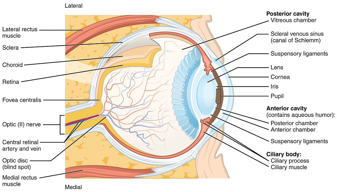
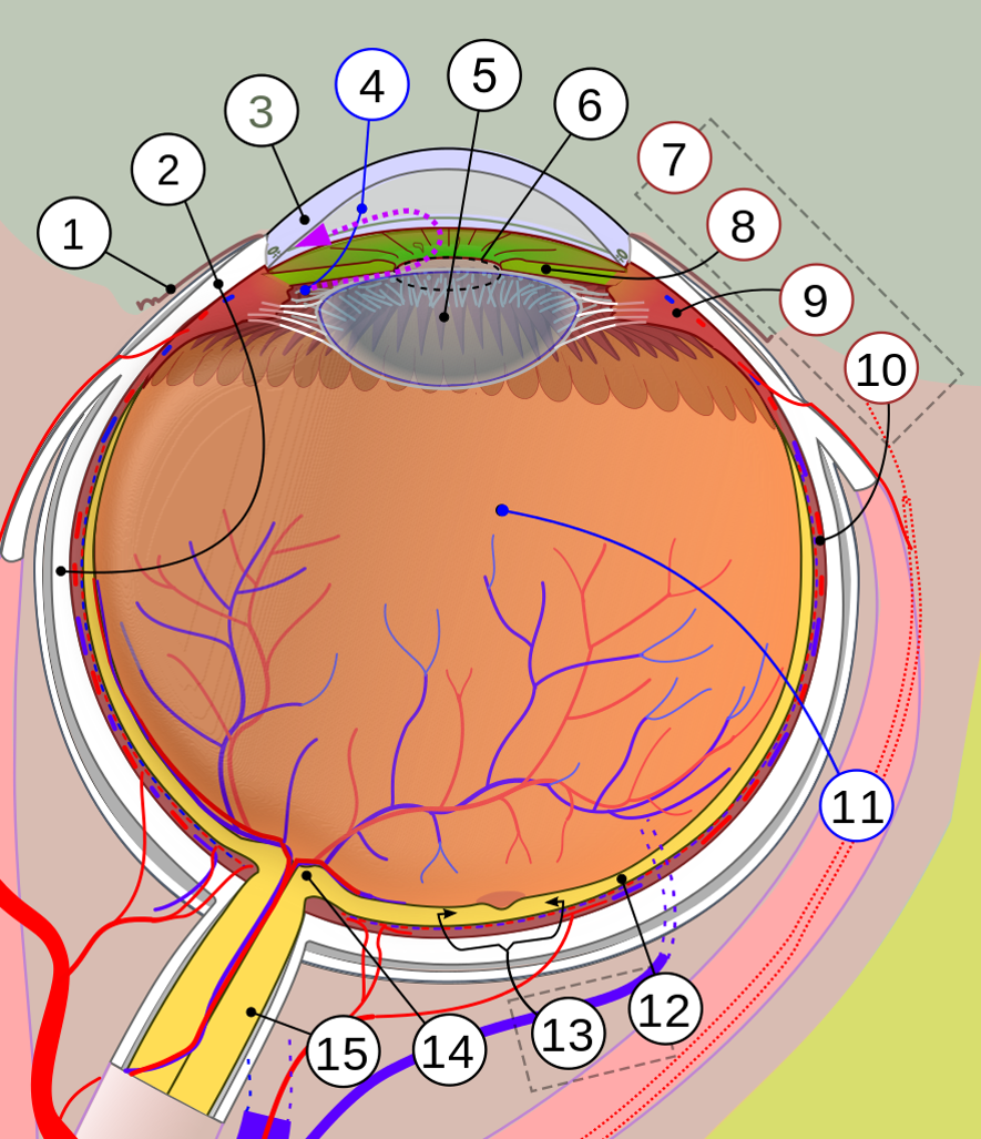
The eye is a hollow, fluid filled organ that is surrounded by three layers of tissue (see image below). The outermost layer, the avascular tunic, is composed of connective tissue. As the name implies there are no blood vessels penetrating this layer. It can be divided into two parts, the sclera, the white part of the eye comprising the posterior 5/6 of the eyeball, and the cornea, the clear window on the anterior surface of the eye. The sclera helps protect the eye and also provides a site of attachment for the six muscles responsible for movement of the eye. The cornea is transparent and functions as the major refractor of the light as it enters the eye. Its transparency is due to the nature of the collagen and proteoglycan fibers that form it. Following are a couple of pictures that help orient us to the anatomy of they eye. It may help to print these and have them in hand as you read the following sections.

Title: File:1413 Structure of the Eye.jpg; Author: OpenStax College; Site: https://commons.wikimedia.org/wiki/File:1413_Structure_of_the_Eye.jpg; License: This file is licensed under the Creative Commons Attribution 3.0 Unported license.

Title: File:Simple diagram of human eye multilingual.svg; Author: Jmarchn; Site: https://commons.wikimedia.org/wiki/File:Simple_diagram_of_human_eye_multilingual.svg; License: This file is licensed under the Creative Commons Attribution-Share Alike 3.0 Unported license.
- Conjunctiva
- Sclera
- Cornea
- Aqueous humour (in anterior and posterior chambers. See purple dotted line)
- Lens
- Pupil
- Uvea with
- Iris
- Ciliary body and
- Choroid
- Vitreous humor
- Retina with
- Macula or macula lutea
- Optic disc → blind spot
- Optic nerve
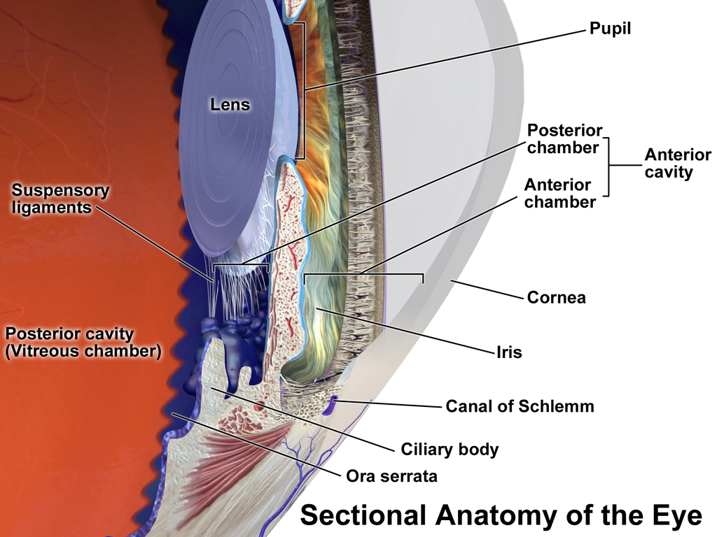
The middle layer of the eye is the vascular tunic. Most of the blood vessels of the eye can be found in this layer. The picture above shows blood vessels of the retina. The blood vessels of the vascular tunic are not shown. If they were shown, you would see them associated with the choroid in the image. The posterior portion of this layer is the choroid. Anteriorly the choroid is continuous with the ciliary body. The ciliary body is composed of a ring of smooth muscle, the ciliary muscle, and the ciliary processes. The ciliary muscle is sphincter-like muscle that is attached to the lens capsule via the suspensory ligaments. It is responsible for adjusting the thickness of the lens. The ciliary processes are secretory structures that produce the aqueous humor that fills the compartment in front of the lens.

Title: Blausen 0390 EyeAnatomy Sectional.png; Author: BruceBlaus; Site: https://commons.wikimedia.org/wiki/File:Blausen_0390_EyeAnatomy_Sectional.png; License: This file is licensed under the Creative Commons Attribution 3.0 Unported license.
The most anterior part of the vascular tunic is the iris. The iris is composed primarily of smooth muscle containing varying amounts of the pigment melanin. The amount of melanin determines eye color, large amounts produce brown eyes, while smaller amounts result in blue or green eyes. The iris is actually two layers of muscle with a circular hole in the center, the pupil. The sphincter pupillae is a circular layer that causes the pupil to constrict (miosis) when it contracts and the dilator pupillae is a radial layer that causes the pupil to dilate (mydriasis) when it contracts (see image below). These layers are innervated by the autonomic nervous system, the dilator is under sympathetic control and the sphincter is under parasympathetic control.

© 2013 Encyclopædia Britannica, Inc. Downloaded from Image Quest Britannica; BYU-Idaho.
Photograph of eye: From left to right we have normal, mydriasis, and miosis.
The innermost layer is the neural tunic or retina. There are actually two distinct layers of the retina. The pigment epithelium is a layer of simple cuboidal epithelium that sits on the choroid. This layer has large amounts of melanin giving it a dark black color. One important function of the pigment retinal is to absorb light that doesn’t strike the photoreceptors and prevent it from being reflected inside the eye. The neural layer is the innermost layer of the wall of the eye and contains the photoreceptors that are stimulated by the entering light. Two distinct anatomical structures on the retina are the optic disk and the fovea centralis. The optic disk, also called the blind spot, is the point where the optic nerve and blood vessels enter the eye. There are no photoreceptors in this area and hence light striking the optic disk cannot be detected. The fovea centralis (fovea = pit) is a small indention located in the center of a special area of the retina called the macula lutea (macula = body, lutea = yellow). The macula is roughly 5 mm in diameter, about the diameter of a pencil eraser, and the fovea is about the size of the head of a pin. When you look at an object the light coming directly from that object focuses on the fovea, it is the portion of the retina with the greatest visual acuity (clarity).
The lens is not technically part of any of these three layers but it is obviously extremely important in focusing light. It is a biconvex structure composed of transparent cells (epithelial cells). These cells have lost their nuclei and other organelles and are filled with transparent proteins called crystallines. It is surrounded by the very elastic lens capsule which, in turn, is attached to the ciliary muscles by the suspensory ligaments. When there is not tension on the suspensory ligaments (ciliary muscles are contracted) the lens assumes its natural shape, this is when it is at its thickest. When the ciliary muscles relax the tension on the suspensory ligaments increases and the lens flattens. Remember the ciliary muscle is a sphincter muscle so when it contracts its diameter decreases, reducing tension on the ligaments attached to the lens capsule.
The lens divides the eye into two fluid filled compartments. The anterior cavity is the space between the lens and the cornea. As was mentioned above, this cavity is filled with the aqueous humor produced by the ciliary processes. Aqueous humor is a watery fluid produced continually and circulates through the cavity before being reabsorbed into the blood. It is important in maintaining proper intraocular pressure as well as circulating nutrients and removing wastes to the cells of the lens and cornea. If the normal circulation is blocked it can result in an inappropriate increase in pressure, a condition known as glaucoma. If not treated, glaucoma can result in vision loss and blindness. You may see some anatomy texts divide the anterior cavity into two “chambers.” The anterior chamber of the anterior cavity would be between the cornea and the iris. The posterior chamber of the anterior cavity would be a very small space between the iris and the lens.
The posterior cavity is the space behind the lens. This compartment is filled with vitreous humor. Vitreous humor is more of a gel, similar to egg white. It also is important in maintaining intraocular pressure, but unlike aqueous humor, turns over very slowly.
Focusing Light on the Retina
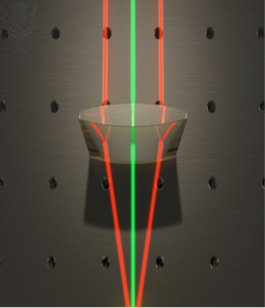
In order to clearly see any object, the first thing that has to happen is that the light reflected off of the object must be focused on the retina. Think of the light being reflected off of a face that you are looking at. Light coming from the subject's nose is spreading out (diverging) as it hits your eye. In order to see the nose clearly, all of the light reflecting from it must be focused on a single spot on the retina. To accomplish this, the light needs to be bent or refracted. You know that light can be focused using something like a magnifying glass. The physics behind this phenomenon has to do with the fact that as light passes through objects of different densities its speed changes. If it strikes the object at a 90 degree angle, even though its speed changes it maintains a straight path. However, if it strikes the object at any other angle (not 90 degrees) it is refracted (see image below). This is why a convex magnifying lens can focus light. We have two convex structures in the eye to bend the light, the cornea and the lens. Also their densities are different than the air and they are different than the aqueous and vitreous humors. We therefore have the ideal conditions for bending the light. However, depending on how close the object is to our eyes the light will be diverging at different angles. The closer it is, the more the light diverges and the more it must be refracted to focus properly. This requires that we be able to adjust the curvature of our refracting surfaces in order to focus properly. The cornea is an excellent refractor, in fact it is responsible for most of the required bending of the light. Its limitation, however, is that it cannot be adjusted. The lens, on the other hand, is adjustable due to its elasticity and the actions of the ciliary muscle. We define a surfaces refracting ability based on its focal point. The focal point is the precise point at which the light rays all converge. As the lens becomes thicker its focal point shortens and it is able to bend light more sharply. On the other had other hand as the lens flattens its focal point becomes longer and it bends the light to a lesser degree. Lets see if we can put this all together and explain we focus objects at different distances from the eye.

© 2013 ENCYCLOPÆDIA BRITANNICA, INC. DOWNLOADED FROM IMAGE QUEST BRITANNICA; BYU-IDAHO.
Illustration of light refraction through a prism which cause the light rays to bend and converge at a single focal point.
We will start with objects that are 20 feet or more from the eye. At this distance the normal eye is designed to focus the object properly without any thickening of the lens. The ciliary muscles would be relaxed and the lens would be at its thinnest. The closest distance at which the lens does not have to thicken for proper focusing is called the far point of vision. For the normal, average eye, the far point of vision is 20 feet. For anything less than this distance from the eye, three things must happen to properly focus the object on the retina. The first we have already discussed; the lens must thicken. This phenomenon is known as accommodation. As objects continue to move closer the lens will thicken more and more until it is at its maximum thickness. If the object is brought even closer it will begin to blur. The closest point that at which we can keep the object in focus is called the near point of vision. The near point of vision changes as we get older. It is only 2-3 inches in infants but may be as far as 5 feet when we get into our late 40’s. The change is due to the fact that the lens becomes less elastic as we age and cannot thicken as much, a condition known as presbyopia.
Focus on the back wall of your room and while maintaining your focus bring your finger in front of your nose a few inches from your face. What do you see? The reason you see two images is because the light is focusing on different parts of the retinas in your two eyes. Now focus on your finger so that you only see one. In order to do that your eyes had to turn in or converge. The closer the object is to the eyes, the more they have to converge. This is the second thing that must happen in order to properly focus on objects less than 20 feet away.
The third thing that happens is that the pupils constrict. The purpose for this constriction is to increase the depth of focus. The depth of focus is how much of the visual field we can keep in sharp focus. Again, place your finger between your face and the computer screen. If you focus on your finger the print on the computer screen will blur and if you focus on the print your finger will blur, we cannot keep both in focus at the same time. Constriction of the pupil increases the depth of focus and helps us keep close objects totally in focus. This is also why we sometimes require additional light to see clearly when doing really close work. Since the pupil is constricted, less light can enter the eye requiring more light to see well.
Focusing Errors
Even though our eyes are designed to focus automatically, like a self-focusing camera, problems do arise. Think of how many people you know who have to wear glasses or contact lenses in order to see clearly. The most common focusing error is near sightedness, or myopia. People who are near sighted can see fine up close but have a difficult time focusing things in the distance. This is usually due to an eyeball that has grown too long. Recall that the eye is designed to properly focus objects greater than 20 feet away without any accommodation of the lens. If the eyeball is too long the image focuses in front of the retina (the focal point is too short) causing objects to appear blurred in our vision. To correct this condition, lenses that spread the light are used to lengthen the focal point and achieve proper focusing. Concave lenses spread light. If you are near sighted and wear glasses, note that the lens of your glasses is thinner in the center than on the edges, creating the concave lens to spread the light. A simple eye test is used to determine if you are nearsighted. This test involves a chart with lines of letters that get progressively smaller as they go down the chart. The subject stands 20 feet from the chart and reads the smallest print that he can. He is then assigned a number based on which line he can read. Normal vision is 20/20 vision. These numbers represent distances. The first number is where the subject is standing, i.e. 20 feet from the chart. The second number is where someone with normal vision would stand to read the same line as the subject. For example if you have 20/20 vision you can see at 20 feet what a “normal” subject would see at 20 feet. If your vision is 20/80 it means that what you see at 20 feet, a “normal” subject would be able to see at 80 feet.
Another vision problem, far sightedness, or hyperopia, is less common and is essentially the opposite of myopia. The subject can see distant objects well but close objects are blurred. The usual cause of this disorder is an eyeball that is too short so the lens cannot thicken enough. Consequently the object focuses behind the retina. To correct far farsightedness, a convex lens is used to shorten the focal length. Children with hyperopia will sometimes grow out of the problem when their eyeball lengthens as they age.
Two other common conditions are astigmatism and presbyopia. Astigmatism is due to an irregularity in the lens or the cornea such that one or both is not symmetrical. This results in part of the object focusing normally while part of it is out of focus. To correct this condition lenses are prepared that are also asymmetrical to counteract the irregularities in the lens or cornea. Presbyopia was alluded to earlier. This is the condition that develops as we age and the lens becomes less elastic. Often, older individuals who have always had normal vision will find that they need reading glasses to help focus the light. The corrective lenses for presbyopia are the same as for far sightedness. They are convex to shorten the focal point of the light.
**You may use the buttons below to go to the next or previous reading in this Module**


