Autonomic Nervous System
Anatomy of the ANS
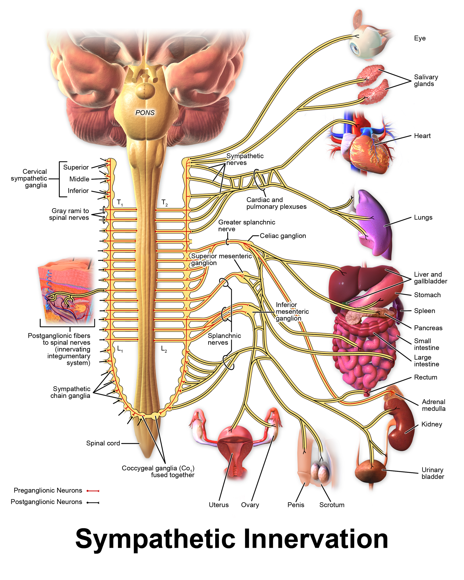
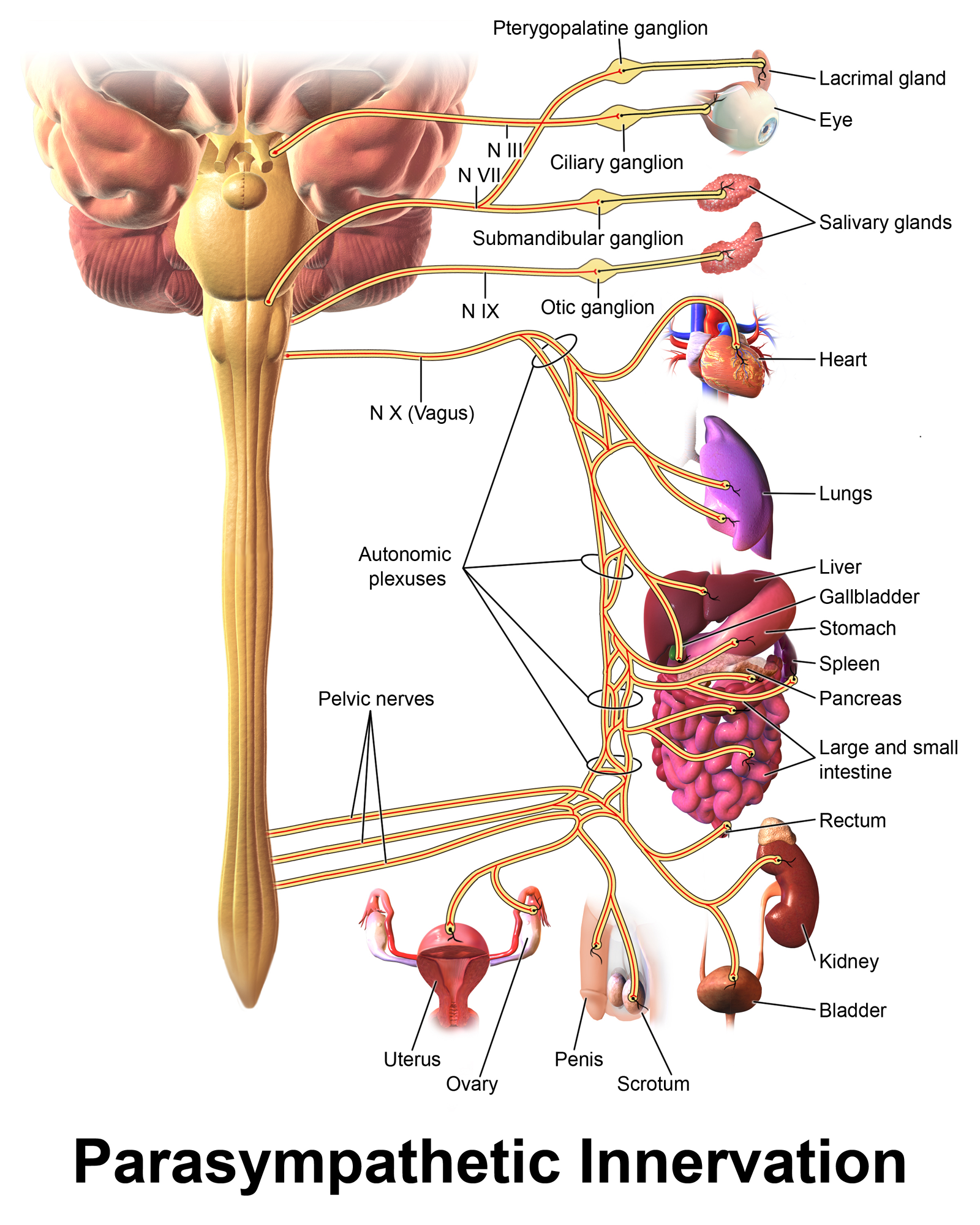
The ANS uses a “two neuron system” to relay electrical signals from the CNS to effectors (organs, glands, and vessels). This is different from the somatic motor division where just one neuron extends from the CNS to skeletal muscle. Parasympathetic and sympathetic divisions are “wired” similarly in that they both have a preganglionic neuron and postganglionic neuron. (Recall from a previous module that a ganglia is a collection of cell bodies located outside the CNS). These neurons get their names from their anatomical location in relation to autonomic ganglia, or relay centers. Autonomic ganglia for the sympathetic division include – sympathetic chain ganglia which are located near the spinal column (labeled at the bottom of the chain in the SNS anatomy image below) and collateral ganglia. The collateral ganglia in the image below are located further away from the spinal column and are labeled Celiac, Superior mesenteric and Inferior mesenteric. Autonomic ganglia for the parasympathetic division are called terminal ganglia and these are located very near the effector they innervate. They are not labeled as there are so many places that parasympathetic neurons synapse with post ganglionic neurons in the very walls of the organs themselves.
The cell bodies for preganglionic neurons are located in either the brainstem or spinal cord, and their axon terminals are located in autonomic ganglia. In the ganglia, a neuron-to-neuron synapse relays information to the cell body of a postganglionic neuron. Postganglionic neurons, also located in the autonomic ganglia, then transmit the signal to effectors. The synapse between the postganglionic neuron and the effector is known as a neuroeffector synapse or neuroeffector junction.

Title: File:Blausen 0838 Sympathetic Innervation.png; Blausen.com staff. "Blausen gallery 2014". Wikiversity Journal of Medicine. DOI:10.15347/wjm/2014.010. ISSN 20018762.; Site: https://commons.wikimedia.org/wiki/File:Blausen_0838_Sympathetic_Innervation.png; License: This file is licensed under the Creative Commons Attribution 3.0 Unported license.
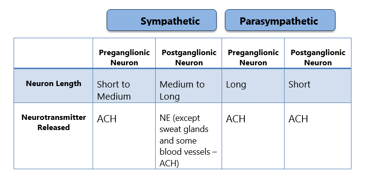
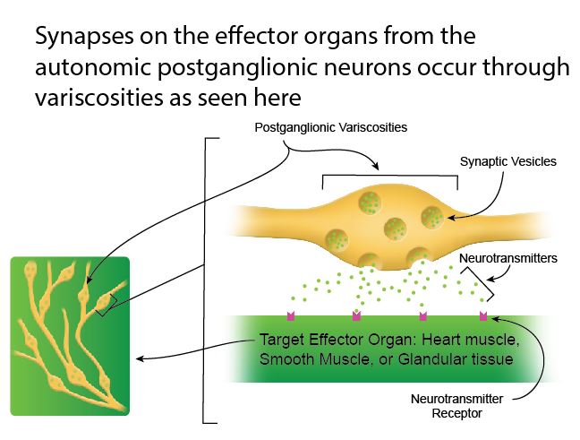
In general, the sympathetic division uses shorter preganglionic neurons and longer postganglionic neurons while the parasympathetic division uses long preganglionic neurons and short postganglionic neurons. Postganglionic release neurotransmitter onto effector organs. However, the synapse is a little unique. Rather than forming a nerve terminal at a synaptic juncition, we see many swellings (or variscosities) develop on the most distal segments of the postganglionic neuron and they secrete neurotransmitter onto the effector tissue (see image below)
Image by BYU-I student Nate Shoemaker Spring 2016

Title: File:Blausen 0703 Parasympathetic Innervation.png; Author: Blausen.com staff. "Blausen gallery 2014". Wikiversity Journal of Medicine. DOI:10.15347/wjm/2014.010. ISSN 20018762.; Site: https://commons.wikimedia.org/wiki/File:Blausen_0703_Parasympathetic_Innervation.png; License: This file is licensed under the Creative Commons Attribution 3.0 Unported license
Compare and Contrast SNS and PNS
The table below helps us compare and contrast some of the characteristics of the SNS and the PNS.

Image by BYU-I student 2013
Ach is short for Acetylcholine and NE is short for Norepinephrine. Acetylcholine and Norepinephrine are neurotransmitters.
**You may use the buttons below to go to the next or previous reading in this Module**


