CELL MEMBRANES-ELECTROPHYSIOLOGY
Refractory Periods
BYUI image: Created F15
Another concept to be discussed is the refractory period. By definition, the refractory period is a period of time during which a cell is incapable of repeating an action potential. In terms of action potentials, it refers to the amount of time it takes for an excitable membrane to be ready to respond to a second stimulus once it returns to a resting state. There are two types of refractory periods; the absolute refractory period, which corresponds to depolarization and repolarization, and the relative refractory period, which corresponds to hyperpolarization. Moreover, the absolute refractory period is the interval of time during which a second action potential cannot be initiated, no matter how large a stimulus is repeatedly applied. The relative refractory period is the interval of time during which a second action potential can be initiated, but initiation will require a greater stimulus than before. Refractory periods are caused by the inactivation gate of the Na+ channel. Once inactivated, the Na+ channel cannot respond to another stimulus until the gates are reset.
Propagation of an Action Potential
Action potentials are usually generated at one end of a neuron and then "propogated" like a wave along the axon towards the opposite end of the neuron.

Title: File:Blausen 0011 ActionPotential Nerve.png; Author: BruceBlaus; Site: https://en.wikipedia.org/wiki/File:Blausen_0011_ActionPotential_Nerve.png; License: This file is licensed under the Creative Commons Attribution 3.0 Unported license.
The image above shows how an action potential might have started near the cell soma and as it propagates down the axon towards the opposite end, the membrane potential behind the moving action potential has repolarized and returned to resting membrane potential. The axon ahead of the current depolarization has not yet depolarized and it is also at resting membrane potential. Where the action potential is occurring we find the membrane potential depolarized and the outside of the membrane at that spot is negatively charged relative to the inside of the membrane at that spot. As sodium rushes in, it will depolarize the next adjacent spot on the axon in the direction that the action potential is propagating. The reason that the action potential does not depolarize the section of axon membrane behind (or in the direction that the action potential just came from) is because that section of membrane is most likely in refractory periods and does not depolarize.

Title: File:Action Potential.gif; Author: Laurentaylorj; Site: https://commons.wikimedia.org/wiki/File:Action_Potential.gif; License: This file is licensed under the Creative Commons Attribution-Share Alike 3.0 Unported license.
The image above is a ".gif" animation and will play only if you see the picture on the internet. As you watch this animation, you will see how an action potential travels as a "depolarization" wave.

BYU-I image: created W15
The image above is another ".gif" animation (must be viewed on the computer and not in print form). This animation shows how an action potential traveling down the axon is similar to stepping on one end of a water balloon. In reality, a pressure wave in the water balloon would get smaller as it traveled down the length, but a traveling action potential (or depolarization wave) is recreated at every spot on the axon that has voltage gated sodium channels to open at threshold. In this way the original strength of the depolarization wave is continually recreated.

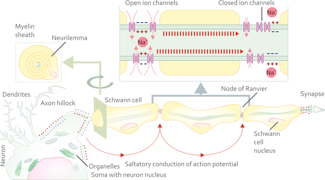
Title: File:Propagation of action potential along myelinated nerve fiber en.png; Author: Helixitta; Site: https://commons.wikimedia.org; /wiki/File:Propagation_of_action_potential_along_myelinated_nerve_fiber_en.png; License: This file is licensed under the Creative Commons Attribution-Share Alike 4.0 International license.
The image above shows myelin on a peripheral nerve axon. The myelin is made up of individual Schwann cells. The myelin covers the axon in a way that "insulates" the axon from depolarization waves. In this way, a depolarization even will occur only at the "Nodes of Ranvier" (or areas of bare axon between individual myelin segments). When a nerve axon is organized in this way with myelin, action potential propagation can travel much faster (nearly 10 times faster than unmyelinated axons).

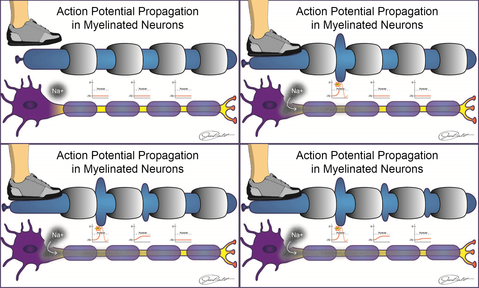
BYU-I image: created W15
The image above is another ".gif" animation. It shows how a myelinated axon might compare to a water balloon with segmented cuffs on it. A pressure wave generated at one segment would travel down the length of the balloon and be recreated at each "node". Notice how the positively charged sodium entering in at the first node causes positive charges to travel down the axon where they can attempt to depolarize each node. The strength of the depolarization wave decreases with distance from the original first depolarization area (just like the pressure wave decreases with distance from the first segment pressed on the water balloon).
The myelinated axon would differ from the balloon in that the original depolarization wave could cause the next node to reach threshold and recreate a depolarization event at the second node that was equal to the first. Consider these two things:
- The original depolarization event can facilitate other nodes in getting closer to threshold.
- Each node that reaches threshold recreates a depolarization wave that is equal to the first
- Depolarization occurs only on bare axon between myelin segments and not along the entire axon surface
These events together make the speed at which and action potential travels to be much faster. This "jumping" of action potential depolarization events from node to node is called saltatory conduction.
Summary
Well, do we have enough information to explain the physiology behind our introductory paragraph? Let's talk about the sense of touch and see if we have a deeper understanding. Consider your fingertips; there are at least 5 different types of touch receptors that allow you to feel various textures and pressures, but how do they work? Touch receptors are really just fancy neurons, but they exhibit the same kinds of phenomena that we just talked about. For example, at rest, they are permeable to K+, but not sodium, so the inside of the membrane is negative relative to the outside. Consequently, Na+ channel proteins are in a closed conformation during rest. In order for us to sense touch we will need to convert the touch stimulus into something the brain can detect; action potentials. The real question is how does touch cause the neuron to send an action potential? Remember that an action potential is caused by Na+ movement across the membrane. Thus, the mechanical action of touch (stimulus) causes a conformation change in a special group of Na+ channels. The action of touch causes them to open, as Na+ moves through those channels, the positive charge of the Na+ ion causes the membrane to change, and other Na+ channels (voltage regulated) respond to the membrane change by opening. This, in turn, causes other channels to open, and the resulting action potential is sent as an electrical current (called an action potential propagation) to the brain. The brain can then interpret the action potentials as physical touch based on where the action potentials originated from. Believe it or not, every external stimulus, whether taste molecules, light waves, sound waves, or mechanical touch, is converted to an action potential. Action potentials are the communications of the body and the brain only works in action potentials.
**You may use the buttons below to go to the previous reading in this Module**


