THE CELL
An Interactive Image that allows you to explore the anatomy of a cell has been added to help you. You can see images of many of the bolded terms below. Be prepared to know where these fundamental cell parts are and what they do.
As technology progressed to the point of peering deeper and deeper into the world of the cell, it became apparent that the cell cytoplasm, when viewed under a light microscope was full of even smaller intracellular structures. The structures are collectively called cellular organelles. This introduction will identify these organelles briefly and subsequent sections will add details.
To help illustrate the function of many of these organelles, let us consider the secretion of insulin by beta cells in the pancreas. In order to secrete insulin, the cell must first make it. This process starts in the cell nucleus . The nucleus houses the genetic material (DNA) of a human cell and provides a location for DNA transcription . Importantly, the nucleus is surrounded by two distinct lipid bilayer membranes. The outer membrane belongs to the endomembrane system (made up of the nuclear envelope, the endoplasmic reticulum, the Golgi apparatus, lysosomes, the plasma membrane, and most vacuoles and vesicles).
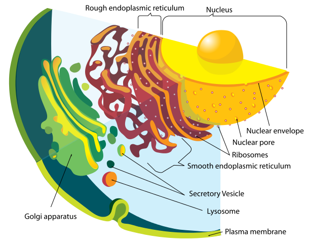
Title: File:Endomembrane system diagram en.svg; Mariana Ruiz LadyofHats; Site: http://en.wikipedia.org/wiki/File:Endomembrane_system_diagram_en.svg; License: Public Domain.
The production and secretion of insulin helps illustrate the coordinated efforts of the organelles in this system. Within the nucleus, the insulin gene (located on chromosome 11) is transcribed from DNA to RNA and then further processed into messenger RNA or mRNA. This mRNA is then transported out of the nucleus to ribosomes docked to the surface of the endoplasmic reticulum (ER). The ER is actually divided into 2 components: the rough ER and the smooth ER. The rough ER is named "rough because it is studded with ribosomes, which create a bumpy surface when viewed under an electron microscope. The function of the ribosome is to perform translation (the conversion of the mRNA into protein). The ribosome is specifically suited to interpret the mRNA nucleotide acid code (a series of adenosines, uracils, guanidines and cytosines abreviated A, U, G, and C respectively) and assign the appropriate amino acid in the creation of a polypeptide chain. This is the first step of making a protein. The process of DNA > RNA > protein is called the central dogma of biology.
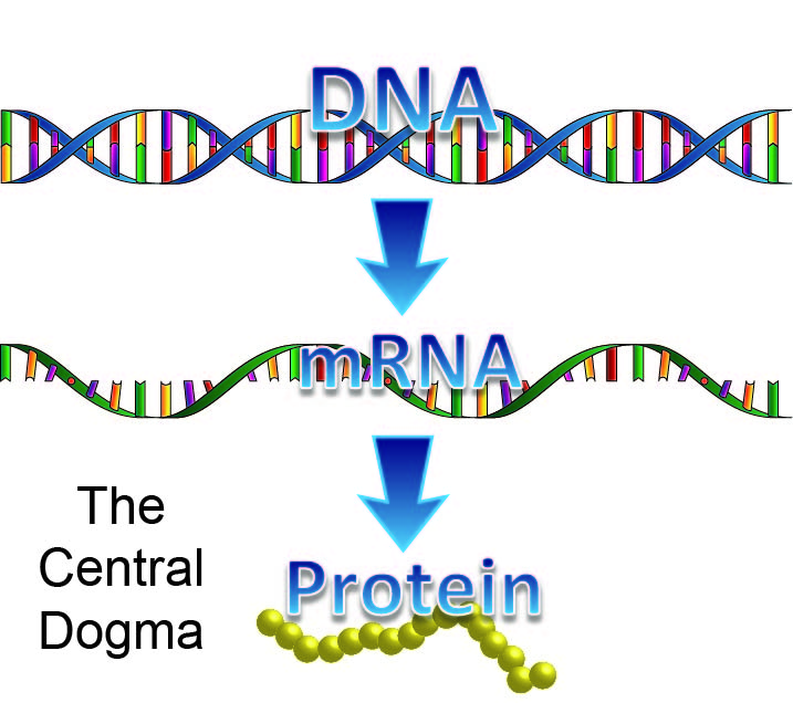
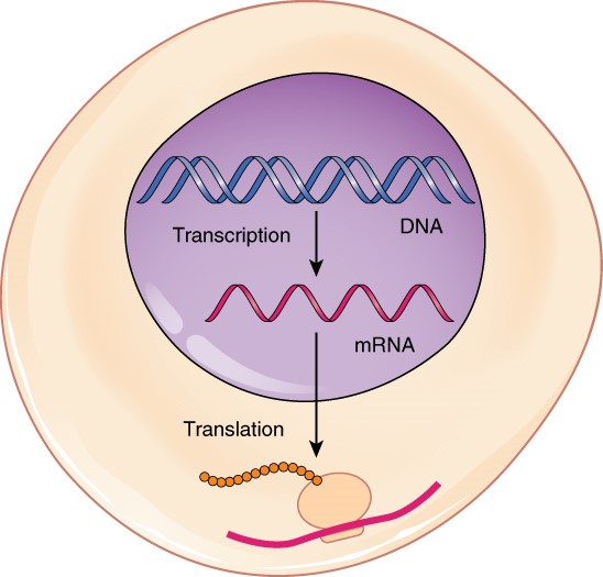
File:0328 Transcription-translation Summary.jpg; Author:OpenStax College; Site:http://commons.wikimedia.org/wiki/File:0328_Transcription-translation_Summary.jpg; License: This file is licensed under the Creative Commons Attribution 3.0 Unported license.
Within the rough ER, the nascent (immature) insulin protein is folded, then transported to the Golgi apparatus . The Golgi apparatus is the location for processing and sorting (think of a giant UPS mail warehouse). Within the Golgi, the nascent insulin is further processed into mature (functional) insulin and packaged into secretory vesicles. These vesicles (now full of insulin) bud off of the Golgi and are transported to the plasma membrane where they await the proper signal for secretion. Secretion occurs as the vesicle fuses with the plasma membrane , expelling its contents into the extracellular space.
Let us now examine the roles of these organelles in greater detail.
The Cell Nucleus
The nucleus is surrounded by a double membrane bound structure that serves to isolate the nuclear contents from the cellular cytoplasm. (This nuclear envelope is dispersed during mitosis as the cell prepares to divide). The outer membrane is continuous with the membranes of the rough endoplasmic reticulum. The inner membrane makes the border to isolate the nucleus. The space between the two membranes is continuous with the space (lumen) inside the endoplasmic reticulum, except at various points where the two membranes are connected by specialized structures known as nuclear pores. The nuclear pores serve as transport pathways between the interior of the nucleus and the cytoplasm. The nucleus contains the genetic material (genes) that are organized into long molecules called DNA that are tightly bound to proteins called histones to form chromatin which is finally organized into chromosomes.
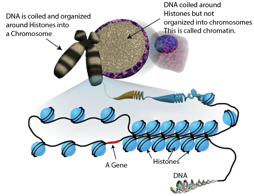
Modified image - Title: File:Sha-Boyer-Fig1-CCBy3.0.jpg; Author: unknown; Site: http://en.wikipedia.org/wiki/File:Sha-Boyer-Fig1-CCBy3.0.jpg; License: This work is licensed under the Creative CommonsAttribution 3.0License.
Gene messages are copied from the DNA as RNA strands, which are further processed into mRNA and sent out of the nucleus through the nuclear pores. The mRNA interacts with ribosomes to produce a specific protein. Many genetic mutations result in errors making the associated proteins non-functional. The nucleus then functions to maintain the integrity of the genes as well as control the turning on or off of the genes. The genes in turn regulate the activity of the cells. Thus, the nucleus is the control center of the cell.
The Endoplasmic Reticulum
As mentioned previously, the endoplasmic reticulum has a rough component and a smooth component. The rough endoplasmic reticulum is associated with ribosomes that constantly bind and unbind to the membrane. Ribosomes bind to the endoplasmic reticulum after they interact with an mRNA strand from the nucleus. The ribosomes "read" the mRNA strand and produce the specific protein associated with the code and secrete it into the lumen of the rough endoplasm reticulum. The newly produced proteins are then folded and prepared for transport to the Golgi complex. The smooth endoplasmic reticulum synthesizes lipids, phospholipids and steroids. In addition, it aids in the breakdown of carbohydrates, and steroids. The membrane contains proteins that move Ca++ into the structure for storage and thus plays and important role in regulating cellular calcium ion concentrations.
The Golgi apparatus
The Golgi apparatus is named after the person that discovered it, an Italian physician named Camillo Golgi who discovered the organelle in 1897. The Golgi apparatus further processes proteins that were first made in the rough endoplasmic reticulum and then distributes the completed proteins to various locations within the cell. The Golgi apparatus is particularly important in the processing of proteins that are destined for secretion outside of the cell. Proteins are sent to the Golgi apparatus from the rough endoplasmic reticulum through transport vessicles that move on the "highway" network of the cell, the cytoskleton (discussed below). The Golgi apparatus is primarily associated with proteins, but also serves in the transport of lipids around the cell and the creation of lysosomes. Perhaps the best analogy for the Golgi apparatus would be that of the post office of the cell.
The Mitochondrion
The mitochondrion (or mitochondria in the plural form) is usually described as the power plant because it generates the energy (in the form of adenosine triphosphate, or ATP) required for normal cellular function. Like the nucleus, mitochondria also have two membranes, which are critical for its function in energy production. (Further detail will be given later as we study metabolism). The mitochondrion is composed of an outer and an inner membrane (a balloon within a balloon) that gives five distinct structural components.
- The outer mitochondrial membrane
- The intermembranous space (the space between the outer and inner membranes)
- The inner mitochondrial membrane
- Cristae (foldings of the inner membrane)
- The matrix (space of the interior of the mitochondrion)
Each region is association with a particular function as it relates to mitochondrial activity. Mitochondria have also been shown to have their own genetic material. The number of mitochondria per cell varies widely with more than 2000 per cell in liver cells down to zero for red blood cells.
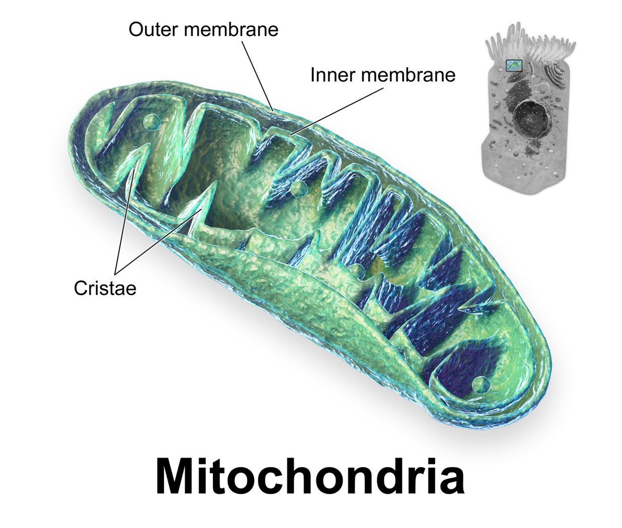
Title: File:Blausen 0644 Mitochondria.png; Author: Blausen.com staff. "Blausen gallery 2014". Wikiversity Journal of Medicine. DOI:10.15347/wjm/2014.010. ISSN 20018762; Site: http://commons.wikimedia.org/wiki/File:Blausen_0644_Mitochondria.png; License: This file is licensed under the Creative Commons Attribution 3.0 Unported license.
Other Organelles
As mentioned, lysosomes are also part of the endomembrane system. Lysosomes are specialized vesicles that bud off of the Golgi apparatus. A lysosome uses a pump within its membrane to transport high concentrations of H+ into its lumen, thus lowering the internal pH. The acidic environment of the lysosome allows it to break down macromolecules (such as proteins). Other organelles involved in recycling used or unneeded materials include proteasomes and peroxisomes . When a cell wants to quickly reduce the amount of a given protein, it can tag that protein with a specific signal (called ubiquitin) that sends that protein to the proteasome for degradation. The peroxisome is responsible for detoxifying harmful substances that may enter the cell.
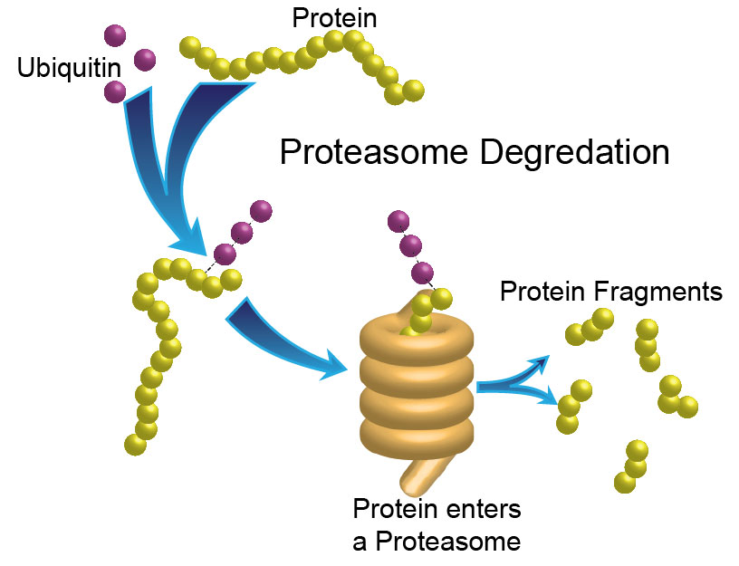
Original image drawn by BYU-I biology department Jan 2015.
The Cytoskeleton
The cytoskeleton, as the name implies, is the structural component of the cell composed of a network of proteins that are constantly destroyed, renewed, and newly built. The cytoskeleton functions in maintaining the cell shape, resisting deformation, movement both inside (transport of vessicles within) and migratory movement, cell signaling, endocytosis and exocytosis, and cell division. The cytoskeleton is composed of three major filaments: microfilaments, intermediate filaments and microtubules filaments. You can explore these components visually at this link:
http://medicineconspectus.blogspot.com/2014/01/cytoskeleton-cell-components-cell.html
Microfilaments are the thinnest of the cellular filaments and are composed of long chains of protein monomers called G-actin. They can generate force by adding monomers that cause the growing strand to push against barriers like the cell membrane. Other proteins like myosin can move along the track and pull against it generating contractile forces in all cells, but which are especially important in muscle cells. Intermediate filaments are stronger than microfilaments and thus help maintain the cell shape. The filaments serve as anchors for other organelles as well as cell to cell junctions. Intermediate filaments are also used in helping to maintain the shape of the nucleus. Microtubules are the largest of all filaments, with a hollow structure made up of protein monomer called tubulin which wind like a spiraling staircase. Microtubules are closely associated with an organizing center called the centrosome . Microtubule networks serve as "highways" for the transport of vesicles, and are important for specialized movements like the swirling tail of sperm cells or the flagellum of bacteria. They also play a crucial role during cell division where they function to pull apart and segregate individual chromosomes.
