CONTROL OF BODY MOVEMENT
VOLUNTARY CONTROL
IntroductionIt is a beautiful, albeit cold, winter evening in Rexburg, Idaho. It just snowed six inches, and your FHE group decided that you could not pass up the opportunity to go sledding at the sand dunes. On the first run of the night your best friend gets huge air on an unseen jump and then lands awkwardly on her back. You quickly sled down to check on her, careful to avoid the jump. When you reach her, she is sitting up but looks confused. You ask her if she is ok and if she remembers what happened, but she just looks at you. Then, without warning, she turns and vomits on the ground. You get the others and decide to take her to the emergency room to get checked out. She is a little wobbly on her feet at first but is able to walk to the car on her own. After waiting in the exam room for a time, the doctor comes in and asks her a series of questions. He then pulls out a mini flashlight and shines it in her eyes one at a time. Curious about why he is shining a light in her eyes, you ask him. He responds that he is evaluating her brain by checking her reflexes. He then has her stand up and walk across the room as he observes. After finishing his exam, he turns to you and says that she has a mild concussion, but she should be fine. He gives you some instructions and warnings, and then you take her home. As you leave, you wonder:
What are reflexes? (You used to think the only way to test reflexes was to hit someone's knee with a hammer!) How do reflexes work? How could the doctor tell that your friend was ok simply by looking in her eyes? What was he looking for as she was walking? How is the nervous system able to control both conscious and unconscious body movements? These are some of the questions that we will attempt to answer in this unit.
Voluntary ControlIn order to understand reflexes and unconscious movement we must first examine how voluntary movements are controlled. Voluntary movements, such as walking upright, are rather complex involving multiple areas within the central (CNS) and peripheral nervous systems (PNS). It is no wonder that it takes time to learn to walk or ride a bike. Once learned, these movements are consciously initiated and then carried out almost automatically. Why are we taught that practice makes perfect? It is because the more we practice a skill the more automatic it becomes. We commonly refer to this phenomenon as "muscle memory."
Such movements depend on upper motor neurons (UMN) and lower motor neurons (LMN). The cell bodies of upper motor neurons are found in the cerebral cortex, where planning, initiation, and coordination of movement occur. Upper motor neurons then synapse with lower motor neurons in cranial nerve nuclei or in the anterior horn of the spinal cord. The lower motor neurons then leave the CNS and synapse with skeletal muscle at the neuromuscular junction. It is this single neuron system from the spinal cord to the muscle that we refer to as the somatic nervous system. To summarize, upper motor neurons initiate movement by sending impulses to lower motor neurons which then relay that information to the skeletal muscle. Thus you can say that voluntary movement comes from the top down and reflexes come from the bottom up. The synapse between the upper motor neuron and the lower motorneuron in the spinal cord is where modulation of both voluntary and reflexive movement takes place.
If needed, look at a helpful picture of the Neuron Pathway.
The lower motor neuron and each skeletal muscle that it innervates is called a motor unit. This has been discussed before, but a quick review would be as follows. A motor unit is a single motor neuron and all the muscle fibers that it innervates. Every time that a motor neuron sends an action potential it will cause an action potential in each of the muscles that it supplies. The size of motor units is quite variable. In areas where we need more precise control each motor neuron innervates very few muscle fibers and in other areas where we need to generate power and precision is not as vital each motor neuron innervates many muscle fibers. Therefore in the places like the eyes and fingers we would have a low muscle fiber to motor neuron ratio whereas in the large muscles of the legs we would have a higher muscle fiber to neuron ratio. The thigh muscles often have a thousand or so fibers per motor unit, while the delicate muscles that move the hand or control eye movement may have only three to five muscle cells per motor unit. The strength of muscle contractions can vary from weak to very strong. For instance, picking up a feather doesn't require much effort (less overall motor units or using motor units with less muscle cells per unit). But, lifting a car would require a much stronger contraction (more motor units and use of units with more muscle cells per unit).
Because muscle fibers contract in an all-or-none fashion the main mechanism for increasing the force of contraction is to stimulate more muscle fibers. This process of increasing the number of motor units activated is called recruitment.
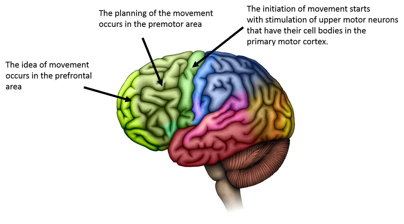
Before a muscle fires, there are several regions of the brain, including the cerebral cortex, basal nuclei (basal ganglia), and cerebellum that work together to control and facilitate the desired movement. We can break this process down into three basic steps: 1) planning, 2) initiation, and 3) execution. The first two steps are mainly controlled by different areas of the cerebral cortex and the last step involves relaying the command from the CNS through the PNS to the muscles involved in the movement.

Image drawn by BYU-I student 2014
The planning step includes forming an idea of what you want to do in the pre-frontal or motor association area and then organizing and coordinating the sequence of events in order to accomplish the movement, which takes place in the premotor area. In the initiation step, action potentials are sent to the upper motor neurons of the primary motor area in the precentral gyrus, which initiates the movement. This signal is sent via action potentials to the lower motor neurons in the cranial nerve nuclei of the brainstem or the anterior horn of the spinal cord. These signals travel from the cerebral cortex down to the brain stem and spinal cord and are thus referred to as descending tracts. The largest of the descending tracts that controls voluntary movement is the lateral corticospinal tract. You have probably heard that the right side of your brain controls the left side of your body and that the left side of your brain controls the right side of your body. This is due to the fact that the upper motor neurons from the lateral corticospinal tract cross over from the right side of the brain to the left side of the spinal cord and then to the left body extremities. This crossing over, called decussation, occurs in a region of the brainstem called the medullary pyramids. The final step is execution of the movement. This takes place as the LMNs send excitatory action potentials to the skeletal muscles responsible for the desired movement, called agonist muscles. They simultaneously send inhibitory action potentials to the antagonist muscles that would oppose the desired movement.
This three-step process that we just explained is much more complex than described above. At each step we are also receiving input from sensory receptors called proprioceptors. They relay information about the position of our body and extremities at any given moment. They also tell us about the direction and rate at which we are moving. We also receive sensory input from our eyes, ears and other sensory organs that we use to modify our movements and react to a dynamic environment. These reactions must take place very quickly to allow adjustments in real time and prevent injury, and thus they function somewhat independently of the higher brain areas. We refer to these reactions as reflexes.
**You may use the buttons below to go to the next or previous reading in this Module**

