Special Senses
THE SENSE OF HEARING: THE COCHLEA
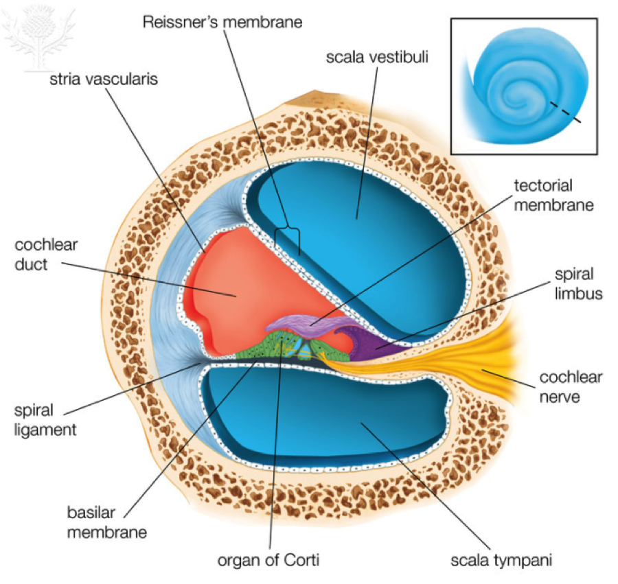
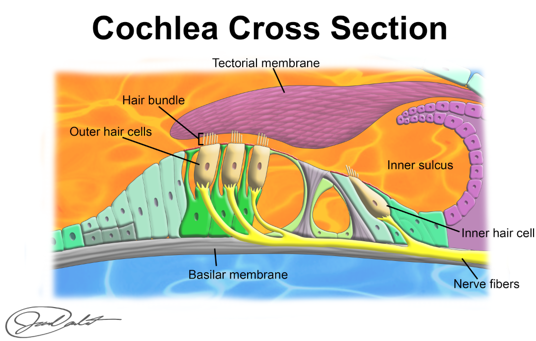
Before we discuss how sound waves are converted to action potentials, we need to understand the structure of the cochlea. This structure gets its name from its shape, cochlea means spiral, or snail shell. The cochlea is a spiral-shaped structure about 3.5 cm long (1.5 inches) that makes 2 ½ turns from top to bottom. It is composed of three parallel chambers that are filled with fluid. The oval window (recall this is a membrane attached to the stapes) communicates with the first chamber, the scala vestibuli, which runs the entire length of the cochlea. When the stapes vibrates it causes the fluids in the scala vestibuli to vibrate. At the very tip of the cochlea, the helicotrema, the scala vestibuli makes a U-turn and becomes the scala tympani. Although they have different names, they are actually one long chamber that folds back on itself. The scala tympani runs parallel to the scala vestibuli and ends at the round window. The round window is a thin membrane between the scala tympani and the middle ear. Thus, when the oval window is pushed in by the stapes, the round window bulges out and when the oval window is pulled out, the round window moves in. It is therefore acting as a pressure release valve, allowing the fluids in these chambers to vibrate (recall that fluids do not compress). The scala vestibuli and scala tympani are filled with perilymph, a fluid that is similar to extracellular fluids. Between these two chambers and also running the length of the cochlea is the cochlear duct. This chamber is filled with endolymph, which unlike the perilymph, resembles intracellular fluid in composition, and thus has a high K+ concentration. Within the cochlear duct is the organ that converts mechanical vibrations to electrical action potentials. This structure is the Organ of Corti or Spiral Organ (see the images below for a cross section of the cochlea and a close up of the spiral organ). The spiral organ sits on the membrane that separates the cochlear duct from the scala tympani, the basilar membrane. As we will explain later, this membrane is responsible for detecting sound waves of different frequencies. Structurally, it is narrow and stiff near the oval window and as it moves toward the helicotrema it becomes wider and more limber. This allows each segment to vibrate at a different frequency. Think of the xylophone you had as a child. The keys on one end were very short and when you struck them they emitted a high pitched sound while the keys at the opposite end were long and emitted a low pitched sound when struck, this is basically the structure of the basilar membrane. Separating the cochlear duct from the scala vestibuli is the vestibular or Reisner's membrane. This is a very flexible membrane that allows the fluid in the cochlear duct to vibrate with the fluid in the scala vestibuli. Located on top of the basilar membrane are four rows of hair cells. There are three outer rows of hair cells and one inner row. These rows run parallel to each other and stretch from the oval window to the helicotrema. As explained later, these are the receptor cells that will generate action potentials. These cells get their name from the rows ofstereocilia on their apical surface. Stereocilia are actually not cilia but instead are more like microvilli. Recall that cilia contain parallel rows of microtubules and are capable of movement whereas microtubules are finger-like projections of the plasma membrane that are supported by microfilaments. In reality, hair cells do have one true cilium called the kinocilium which is adjacent to the longest microtubule. Interestingly, in mammalian cochlea, these kinocilium disappear shortly after birth and no one knows what their function is. Each hair cell has 50-150 stereocila of different lengths. They are arranged much like the reception bars on your cell phone, gradually increasing in length from one side of the cell to the other. At the point where the stereocilium attaches to the rest of the cell its diameter is much smaller, creating a hinge-like structure that allows it to bend back and forth. Located on these stereocilia are the mechanically-gated ion channels that will respond to the vibrations of the basilar membrane. Just above the hair cells is another structure called the tectorial membrane. It extends like a shelf over the hair cells and the longest stereocilia in the outer three rows of hair cells are imbedded in this membrane.

© 2013 Encyclopædia Britannica, Inc. Downloaded from Image Quest Britannica; BYU-Idaho.
Cross sectional diagram of the cochlea illustrating the three chambers and the organ of corti.

Produced by BYU-Idaho studnet Jared Cardinet F14
Diagram of the organ of corti and the associated hair cells and neurons.
Conversion of Vibrations in the Air to Perceived Sounds
Transfer of Vibrations in Air to Vibrations in Fluids: The first challenge that our ears face is transferring the vibrations in the air, to vibrations in a fluid. Because the density of the fluid in the inner ear is much greater than the density of air it requires more energy to generate sound waves in the fluid than in the air. Think of being underwater at a swimming pool and listening to people talk, it is very hard to hear and understand. It is the middle ear's responsibility to amplify the sound waves so that their energy is not lost. This is accomplished in two ways. First, the arrangement of the ear ossicles amplifies the sound. Second, and probably more importantly, the tympanic membrane has about 20 times more surface area than the oval window. This size difference results in concentrating the energy on the oval window. Think of how you might move a large rock with a pry bar. You would place the fulcrum close to the stone to gain the maximal mechanical advantage of the bar. the long end of the bar would be analogous to the tympanic membrane and the short end would be analogous to the oval window. These mechanisms are so effective that very little, if any, energy is lost as it is transferred from air waves in the external ear to fluid waves in the internal ear.
Detection of Sound Waves of Different Frequencies: As explained earlier, sound waves of different frequencies are perceived as different pitches. Therefore, the inner ear needs a way of detecting the different frequencies. The structure in the inner ear tasked with this responsibility is the basilar membrane. Recall the design of the basilar membrane, it is narrow and stiff near the oval window and gradually gets wider and more limber as it progresses toward the helicotrema. Think of the example of the xylophone mentioned earlier. When you strike a key on a xylophone it always sounds the same because it always vibrates at the same frequency. Another analogy might be a guitar string. As you tighten a guitar string making it stiffer, it vibrates at a faster rate and produces a sound of a higher pitch. Also on the guitar as you shorten the string by pressing on a fret with your finger the pitch gets higher. At a given tension and length the guitar string always vibrates at the same rate so we always perceive it as the same pitch. The basilar membrane functions in much the same way. Each segment of the membrane has an innate frequency. If it were a guitar string and you plucked it at certain point along its length it would always vibrate at the same rate at that point. A different point on the basilar membrane would vibrate at a different rate. When a vibration in the fluid reaches the segment of the basilar membrane that has the same innate frequency, it will cause the basilar membrane to vibrate. This phenomenon is known as resonance. Based on this principle of resonance the basilar membrane is able to respond to all of the different frequencies in the sounds we hear, within the range of human hearing.
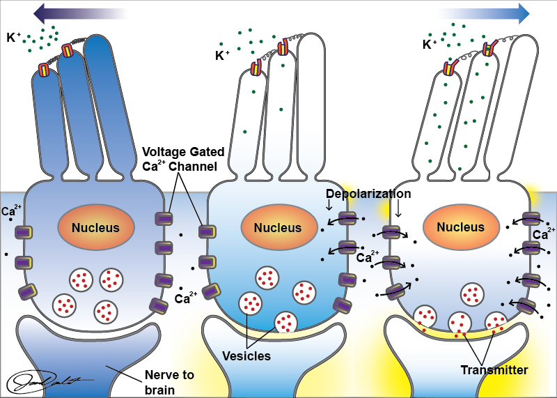
Conversion of a Sound Wave to an Action Potential: The function of any sensory organ is to convert a sensory stimulus to an action potential that can then be transmitted to the brain. In this case, the sensory signal is the sound wave. The responsibility of converting vibrations into action potentials falls upon the inner row of hair cells in the cochlea. Recall that the apical end of the hair cell contains the stereocilia and that they are arranged in order of ascending lengths from one side of the cell to the other. The membranes of the stereocilia contain mechanically gated cation channels. Extending from the gate of the ion channel to the adjacent, taller, stereocilium is a fibrous protein called a tip link (see image below). When the stereocilia bend toward the longest stereocilium the tension in the tip link increases, pulling the gates on the ion channels open, and when they bend in the opposite direction the tension decreases and the gates close. The stereocilia are bathed in the endolymph of the cochlear duct. Endolymph is similar to intracellular fluid and has a high K+ concentration. When the gates on the cation channels open, K+rushes into the cell, depolarizing the membrane. This depolarization opens voltage gated Ca2+ channels on the basal membrane of the hair cell allowing Ca2+ to enter. The influx of Ca2+ stimulates the release of neurotransmitter by the hair cell triggering an action potential in the neuron that synapses with the hair cell. The axons of these neurons form the cochlear nerve that transmits the action potential to the auditory cortex of the brain. In hair cells at rest, about 10% of the K+ ion channels are open resulting in a low frequency of action potentials traveling to the brain when it is perfectly quiet. This allows for both an increase in action potential frequency when hair cells bend toward the longest stereocilium, and a decrease in frequency of action potentials when the hair cells bend the other way (see image below).

Produced by BYU-Idaho studnet Jared Cardinet F14
Hair Cells of the Spiral Organ
Perception of Sound: Once the action potential is generated and sent to the brain it is the function of the auditory cortex to convert that action potential into a perception. Each region of the cochlea is hardwired to its own specific region of the auditory cortex. When that particular region of the brain receives input from the ear we perceive the unique pitch associated with that frequency of sound wave. It's kind of like a piano where each key is like a different segment of the cochlea. That key is linked to a specific string in the piano such that each time the key is struck we hear the same sound. In this case the strings would be like a specific region in the auditory cortex. Each time an action potential reaches that specific segment of the auditory cortex we perceive the same sound. Therefore, pitch is determined by the region of the brain that receives input from the cochlea. Loudness, on the other hand, is determined by the number of action potentials that reach the brain. Recall that the loudness of a sound is a function of the amplitude of the sound wave. Sound waves of higher amplitude cause the hair cells to vibrate more vigorously, which would cause more ion channels to open. This would result in a greater depolarization of the hair cell, more Ca2+ entry through the voltage-gated ion channels and more neurotransmitter release. The end result is a greater frequency of action potentials going to the auditory cortex, which is perceived as a louder sound. A common misconception is to equate the frequency of action potentials with the frequency of the sound waves. The frequency of action potentials is a function of the amplitude of the sound wave whereas the frequency of the sound waves determines which portion of the auditory cortex receives the action potentials.
Other Factors Influencing Our Perception of Sound: There are several other factors that impact what we hear. An important consideration is the function of the three outer rows of hair cells. About 90% of the neurons of the cochlear nerve arise from the inner row of hair cells and these are thought to be key to communicating with the auditory cortex. The outer hair cells, on the other hand, are implicated in a process called cochlear amplification. These hair cells have special proteins in their plasma membranes that allow the cell to actively lengthen and shorten. This action can either enhance or reduce the movement of specific regions in the cochlea. The outcome of this action is thought to help focus the sound to specific regions of the spiral organ so that we can better detect the different frequencies of sound waves. Also recall that only the stereocilia of the outer three rows of hair cells are inbedded in the tectorial membrane, those of the inner hair cells are not. What then causes the stereocilia in the inner hair cells to bend? The structure of the spiral organ allows the basilar membrane and the tectorial membrane to function like a bellows. The two membranes form a pocket, the inner sulcus, behind the inner row of hair cells. When the basilar membrane moves up it squeezes the bellows and endolymph flows out of the inner sulcus bending the stereocilia one way and when the basilar membrane moves down the bellows enlarges pulling endolymph into the inner sulcus bending the stereocilia the opposite way.
Hearing loss
There are three forms of hearing loss: conductive, central, and sensorineural. Conductive hearing loss is a result of sound waves being unable to move from the external ear to the inner ear. This can be caused by a plugged ear canal (excessive ear wax), infection of the middle ear or calcification of the stapes to the oval window. Anything that prevents conduction of sound through the external ear or proper vibration of the middle ear bones is termed conductive hearing loss. Central hearing loss results from damage of the auditory cortex, usually caused by a stroke. Sensorineural hearing loss is caused by damaged to the structures of the inner ear (hair cells, cochlear neurons, viscous fluid). A common cause of sensorineural hearing loss is exposure to loud sounds. In humans, this loss is irreversible at present. Interestingly, birds have the ability to regenerate hair cells after complete destruction. Study of hair cells in birds may one day lead to the ability to replace damaged hair cells in humans.
**You may use the buttons below to go to the next or previous reading in this Module**


