CONTROL OF BODY MOVEMENT
SOMATIC REFLEXES
In our discussion we will examine four major reflexes that are integrated within the spinal cord: the stretch reflex, the Golgi tendon reflex, the withdrawal reflex and the crossed extensor reflex. Although each of these reflexes is integrated within the spinal cord, they can be influenced or modified by higher brain centers to either exaggerate or suppress the response. Somatic reflexes involve specialized sensory receptors called proprioceptors that monitor the position of our limbs in space, body movement, and the amount of strain on our musculoskeletal system. The effectors involved in these reflexes are located within skeletal muscle.
Stretch Reflex
Think back to the last time you had a sports physical or a routine physical examination. Why did the doctor tap your leg just below the knee? What information can he possibly gather from this simple procedure? The magic of examining reflexes comes from the phenomenon that, under normal circumstances, a specific stimulus will elicit a predictable response. In the case of the knee-jerk reflex the expected response is extension of the leg at the knee. If the reflex is greater than expected (hyperactive), less than expected (hypoactive) or totally absent, that suggests potential pathology. Now let's look at how the stretch reflex works.
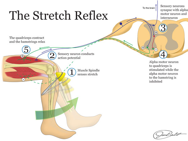
Muscle spindles are specialized proprioceptors that monitor muscle length. They are bundles of modified skeletal muscle fibers with extensive sensory and motor innervation. These fibers, called intrafusal fibers, run parallel to the contractile skeletal muscle fibers called extrafusal fibers that make up the bulk of skeletal muscle. Muscle spindles are scattered throughout skeletal muscle, but they occur in the highest density near tendinous insertions and in muscles involved in fine motor control (i.e. the small muscles of the hand etc). Intrafusal fibers are only capable of contraction at their tapered ends where they are innervated by gamma motor neurons. (The contraction is too weak to contribute to gross movement but is important in maintaining the sensitivity of the muscle spindle while the muscle is either shortened or lengthened.) Sensory neurons innervate the noncontractile central region of the intrafusal fibers. If stretched, the sensory fiber associated with the muscle spindle will be activated and result in stimulation of an alpha motor neuron (a type of lower motor neuron) in the anterior horn of the spinal cord. The alpha motor neurons directly innervate the skeletal muscle where the muscle spindle is located. This is an example of a monosynaptic reflex because the sensory neuron synapses directly with the motor neuron and occurs without any input from the upper motor neuron.
Imagine stepping out of the driver's seat of your car onto a patch of ice in the parking lot. As your weight transfers to your left foot and starts to slide out from under you, what happens? The muscle spindles in your left inner thigh (adductors) are quickly stretched and send a message to your alpha motor neurons in the spinal cord begging for help. The alpha motor neurons then cause contraction of the same inner thigh muscles (adductors) that were stretched, and you narrowly avoid the pain of a groin injury. All of this happens so fast (signals are sent at speeds around 350 miles per hour) that you have already recovered by the time you are aware that you were in trouble. When a muscle is stretched, the muscle spindles are stimulated and thus increase the frequency of action potentials sent to the lower motor neurons in the CNS. The increased action potential frequency causes alpha motor neurons to rapidly fire, resulting in muscle shortening. This reflexive contraction, in the direction directly opposite to the initial stretch, protects skeletal muscle from damage due to overstretching.

Image drawn by BYU-I student Jared Cardinet Winter 2015
The same process that we described above also relates to other very common situations. For example, as you are reading this you may be experiencing some drowsiness. We will assume that is because you have stayed up way too late! As you get tired you may have experienced the feeling of nodding off, where your head starts to fall forward followed by an almost violent jerking motion as you bring your head upright again. Your muscle spindles are key in maintaining posture, whether we are talking about nodding off in class or whether we are talking about staying upright as you walk down the street.
So, now that the muscle that was being stretched is shortened, what happens to the muscle spindle? Does it become insensitive to further changes in that muscle's length? Remember, we said that gamma motor neurons innervate the contractile ends of the muscle spindle. As the alpha motor neurons activate extrafusal fibers, causing shortening of the muscle, gamma motor neurons activate the muscle spindle. We refer to this as alpha-gamma co-activation. This causes the tapered ends to contract, thus maintaining a baseline tension on the central region of the muscle spindle that is sensitive to stretch. It is in this manner that the muscle spindle is able to maintain its sensitivity through a wide range of muscle length.
In fact, even when a muscle is at rest the muscle spindle sends out a relatively steady stream of action potentials which helps to maintain a low level of muscle activity. This constant tension of the muscle is what we refer to as muscle tone.
Up to this point we have only addressed activation of the muscle group that is being stretched. This is important but body movement is controlled by opposing muscle groups, the agonist and antagonist muscles. The agonist muscle is the muscle that contracts to cause a certain movement to happen and the antagonist is the muscle group that would do the opposite action. In the example of the knee jerk reflex the quadriceps would be the agonist and the hamstring would be the antagonist. In order to extend the leg at the knee we must contract the quadriceps, which we do via activation of the alpha motor neurons, but we must also relax, or inhibit, the hamstring. We accomplish this through a phenomenon called reciprocal inhibition. The sensory neuron that synapses with and excites alpha motor neurons supplying the quadriceps also synapses with an inhibitory interneuron. The inhibitory interneuron effectively shuts down the alpha motor neurons to the hamstring. This allows the leg to extend at the knee.
Golgi Tendon Organ (GTO)
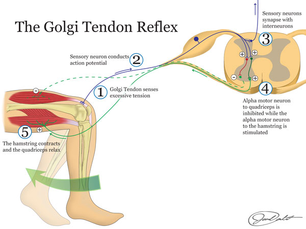
Whereas muscle spindles respond to stretch another type of sensory system responds to tension. You might think that stretch and tension are pretty much the same thing but they are not. Have you ever tried tying your shoes really tight and as you are pulling on the laces, which increases tension, one of the laces snaps? It is pretty inconvenient when you have to replace a shoelace but think if that was your muscle! At times our muscles are capable of generating sufficient power to damage tendons or even break bones. They can cause avulsion, where the tendon tears off a piece of the bone at its attachment site. In order to prevent this we have a safety mechanism in place called the Golgi tendon organ. Where we could consider the stretch reflex to be excitatory and cause contraction of the stretched muscle group the Golgi tendon reflex would be considered inhibitory and causes relaxation of the affected muscle. Therefore the result of activation of a GTO would be the opposite of the activation of a muscle spindle. The main purpose of GTOs is to prevent excessive tension on tendons and thus prevents injury.
Golgi tendon organs are composed of encapsulated nerve endings that are found interwoven with collagen fibers near the transition from muscle to tendon. These nerve endings monitor tension on the tendon rather than muscle length as muscle spindles do. As a muscle contracts it develops tension on the tendon which is detected by the GTO. The GTO then sends action potentials, via afferent neurons, to the dorsal horn of the spinal cord where they synapse with inhibitory interneurons. The interneuron then synapses with and inhibits the alpha motor neurons in the anterior horn of the spinal cord. Inhibition of alpha motor neurons will effectively shut off the "power" to the muscle causing it to relax. You can think of this phenomenon almost like a circuit breaker. If there is a spike in power coming into your home that could potentially damage electrical devices the circuit breaker is tripped, temporarily shutting off electricity to those electrical devices.

Image drawn by BYU-I student Jared Cardinet Winter 2015
You might ask yourself, "If this prevents excessive tension on muscles, what about those stories I have heard about mothers lifting cars off of babies and such?" Well, remember that this is a reflex and is generally managed from the bottom up without too much oversight from the upper motor neurons. In some circumstances, such as the super human feats of strength you have heard about, the CNS has the ability to override the reflex of the GTO. This happens as upper motor neurons modify the reflex at the level of the spinal cord. This allows extreme amounts of force and tension to be achieved, but the downside is that it usually causes pretty severe damage to the musculoskeletal system.
Withdrawal Reflex and Reciprocal Inhibition

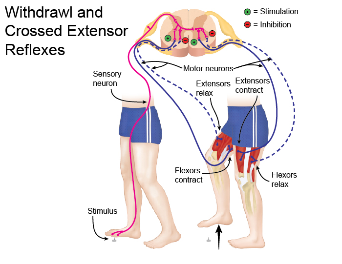
Image drawn by BYU-I student Nate Shoemaker Spring 2016
Have you ever stepped on something sharp with your bare feet or touched something hot with your hand? If the answer is yes then you have experienced the grace of the withdrawl reflex. If the answer is no, you need to live a little! The withdrawl reflex is yet another way that we are hard wired to avoid pain and tissue damage. We have free nerve endings, called nociceptors, scattered throughout our body that are sensitive to pain. When stimulated these sensory neurons activate lower motor neurons in the spinal cord. The lower motor neurons then stimulate contraction of skeletal muscle to remove or withdraw ourselves from the pain generator. In general, this will take place as flexor muscles are stimulated to contract, such as the hamstrings and hip flexors if you step on a tack or the biceps when you touch a hot stove. For this reason the withdrawl reflex is sometimes called the flexor reflex.
In order for this to happen efficiently, we need to stimulate the flexor muscles and at the same time inhibit the extensor muscles. This phenomenon, called reciprocal inhibition, that was discussed in terms of the knee-jerk reflex is also at play here. The pain neuron, as it enters the dorsal horn of the spinal cord, will branch to stimulate an excitatory interneuron and an inhibitory interneuron. The excitatory interneuron then stimulates muscle contraction of the flexor muscle while the inhibitory interneuron causes the antagonist muscle, or the extensors to relax.
Crossed Extensor Reflex
The crossed extensor reflex is yet another way that your body protects itself. When you step on that tack and reflexively pull your foot away you quickly find yourself supporting all of your weight on one leg. Without the crossed extensor reflex, instead of standing on one leg after stepping on a tack you would probably wind up on your backside.
Again, when you step on a tack and stimulate the pain fibers in your foot they send signals to the spinal cord through the dorsal horn. In addition to sending branches to excitatory and inhibitory interneurons on the same side of the body the pain neuron also sends a branch to an excitatory interneuron that crosses over to the opposite side of the spinal cord and stimulates a lower motor neuron. This lower motor neuron stimulates the extensor muscles on the opposite side of the body in preparation for the increased load as you shift your weight to that side.
Summary
Now that you know what reflexes are and how they work, let's revisit the question, "How could the doctor tell that your friend was ok simply by looking in her eyes?" The answer is that he was checking the integrity of a different reflex, the pupillary light reflex. Remember, the clinical usefulness of checking reflexes is that specific stimuli should elicit predictable responses. Therefore, you would anticipate that shining a bright light in a person's eyes would cause the pupils to constrict, and that is exactly what should happen, but how? There are special receptors in the eye that are sensitive to light. When stimulated, like when the doctor shined the bright light in your roommate's eye, they transmit signals through the optic nerve to the midbrain. In the midbrain these neurons stimulate the occulomotor nerves, which supply the muscles that cause constriction of the pupil. Thus by checking the pupillary light reflex the physician was able to quickly evaluate the seriousness of the injury. In the case of severe brain injury this reflex can be compromised so that the bright light would not cause the anticipated pupil constriction.
Some nice Youtube summaries of these reflexes are found at: http://www.youtube.com/user/HAPProf?feature=watch
**You may use the buttons below to go to the next or previous reading in this Module**

