INTRODUCTION TO THE NERVOUS SYSTEM
NEURON STRUCTURE AND CLASSIFICATION
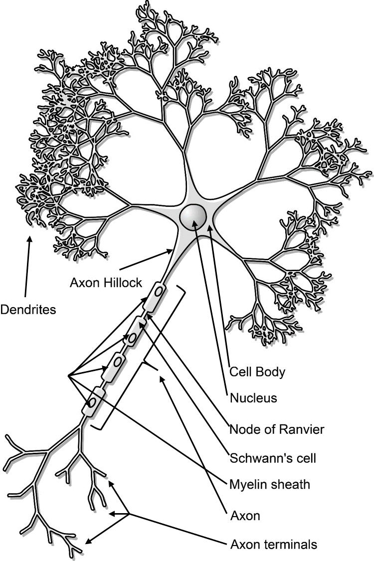
Neurons have four specialized structures that allow for the sending and receiving of information: the cell body (soma), dendrites, axon and axon terminals (see lowest figure).
Cell body or soma: The cell body is the portion of the cell that surrounds the nucleus and plays a major role in synthesizing proteins.
Dendrites: Dendrites are short, branched processes that extend from the cell body. Dendrites function to receive information, and do so through numerous receptors located in their membranes that bind to chemicals, called neurotransmitters.
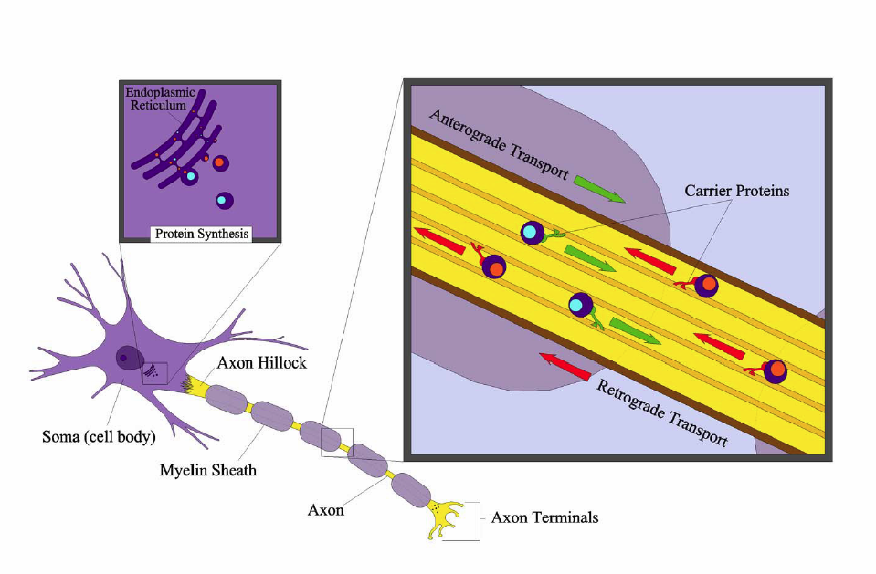
Axon: An axon is a large process that extends from the cell body at a point of origin-called the axon hillock-and functions to send information. In contrast to the shorter dendrites, the axon can extend for more than a meter. Because of this length, the axon contains microtubules and is surrounded by myelin. Microtubules are arranged inside the axon as parallel arrays of long strands that act as highways for the movement of materials to and from the soma. Specialized motor proteins "walk" along the microtubules, carrying material away from the soma (anterograde transport) or back to the soma (retrograde transport). This system can move materials down the axon at rates of 400mm/day (see lowest figure). Myelin consists of totally separate cells that coil and wrap their membranes around the outside of the axon. These are essential for electrical insulation and to speed up action potential propagation.

Image produced by BYU-Idaho Student Jared Cardinet 2013
Image shows Anterograde and Retrograde transport in an axon.
Axon terminals: Once an axon reaches a target, it terminates into multiple endings, called axon terminals. The axon terminal is designed to convert the electrical signal into a chemical signal in a process called synaptic transmission (further explained in the section "Physiology of the Neuron").
Most neurons are amitotic or lose their ability to divide. Exceptions to this rule are found in olfactory neurons (those associated with smell) and hippocampal regions of the brain. Fortunately, lifespans of amitotic neurons is near 100 years. Still, if a neuron is damaged or lost, it is not easily replaced. For this reason, there is usually limited recovery from serious brain or spinal cord injuries. Perhaps the slow recovery rate or lack of regeneration is to ensure that learned behavior and memories are preserved throughout life. Neurons also have exceptionally high metabolic rates and subsequently require high levels of glucose and oxygen. The body will go to great lengths to ensure that neurons are adequately fed; in fact, if for some reason the brain detects that it is not receiving adequate amounts of nutrition, the body will shut down immediately (i.e., faint).

Title: Neuron-figure-notext.svg; Author: Nicolas.Rougier; Site: https://commons.wikimedia.org/wiki/File:Neuron-figure-notext.svg; License: This file is licensed under the Creative Commons Attribution-Share Alike 3.0 Unported license.
Illustration of key neuronal structures
Classification of Neurons
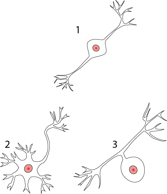
Structural classification of neurons is based upon the number of processes that extend out from the cell body. Three major groups arise from this classification: multipolar, bipolar, and unipolar neurons.
Multipolar neurons are defined as having three or more processes that extend out from the cell body. They comprise of more than 99% of the neurons in humans, and are the major neuron type found in the CNS and the efferent division of the PNS.

Title: Neurons uni bi multi pseudouni.svg; Author: Pseudounipolar_bipolar_neurons.svg: Juoj8 derivative work: Jonathan Haas; Site: https://commons.wikimedia.org/wiki/File:Neurons_uni_bi_multi_pseudouni.svg; License: This file is licensed under the Creative Commons Attribution-Share Alike 3.0 Unported license.
Structural classification of neurons. 1) Bipolar; 2) Multipolar and 3) Unipolar.
Bipolar neurons have only two processes that extend in opposite directions from the cell body. One process is called a dendrite, and another process is called the axon. Although rare, these are found in the retina of the eye and the olfactory system.
Unipolar neurons have a single, short process that extends from the cell body and then branches into two more processes that extend in opposite directions. The process that extends peripherally is known as the peripheral process and is associated with sensory reception. The process that extends toward the CNS is the central process. Unipolar neurons are found primarily in the afferent division of the PNS.
Functional Classification of Neurons
Neurons are classified functionally according to the direction in which the signal travels, in relation to the CNS. This classification also results in three different types of neurons: sensory neurons, motor neurons, and interneurons.
Sensory neurons, or afferent neurons transmit information from sensory receptors in the skin, or the internal organs toward the CNS for processing. Almost all sensory neurons are unipolar.
Motor, or efferent neurons transmit information away from the CNS toward some type of effector. Motor neurons are typically multipolar.
Interneurons are located between motor and sensory pathways and are highly involved in signal integration. The vast majority of interneurons are confined within the CNS.
**You may use the buttons below to go to the next or previous reading in this Module**


