INTRODUCTION TO THE NERVOUS SYSTEM
GLIAL CELLS
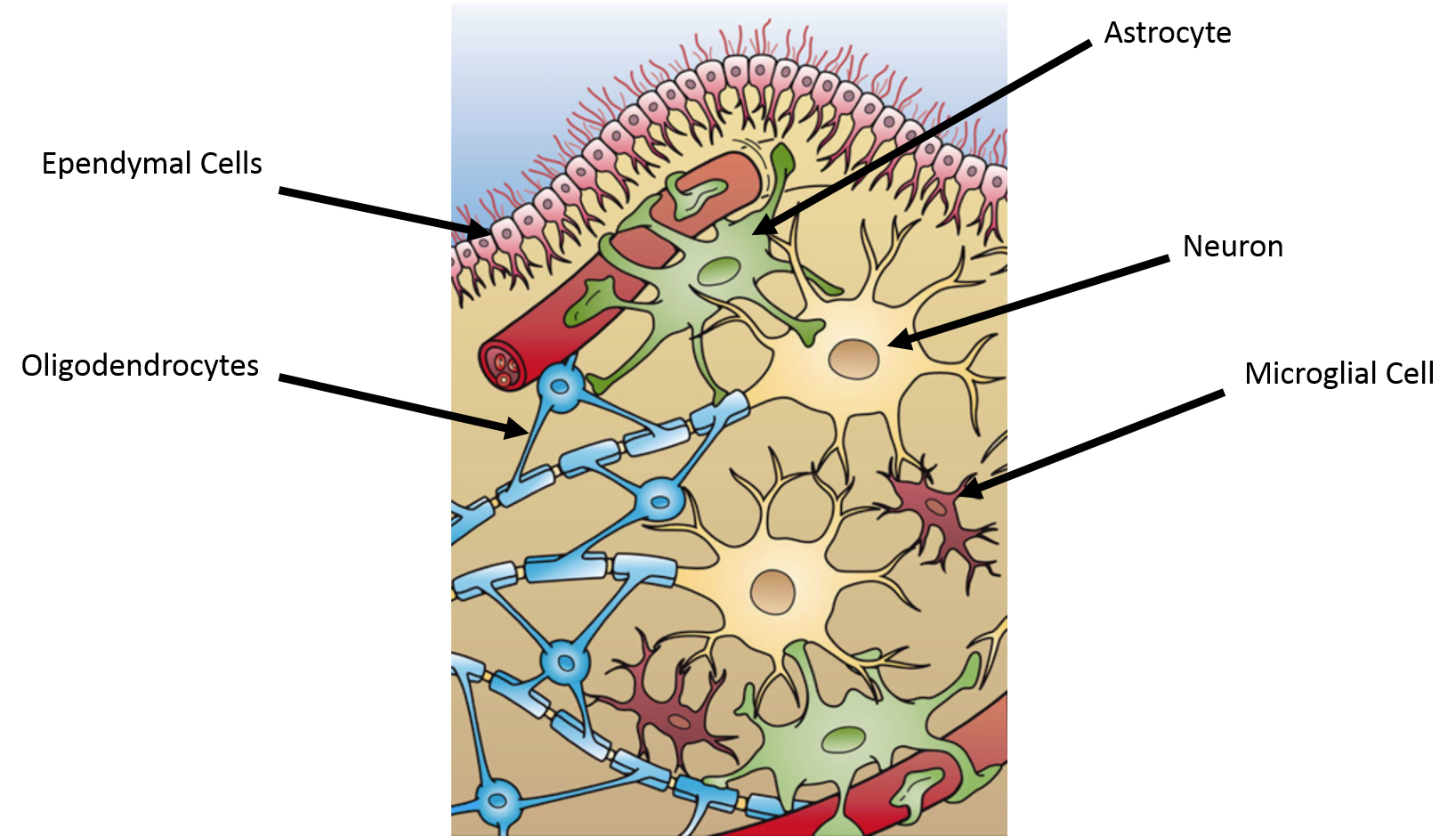
Unlike neurons, the glial cells can be replaced if they are damaged. Glial cells compose half of the volume of the brain and are more numerous than neurons. There are four major types of glial cells in the CNS: the astrocyte, the oligodendrocyte, the ependymal, and the microglial cell.
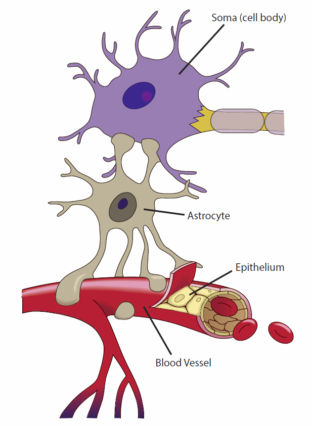
The astrocyte: Astrocytes have an enormous amount of processes that wrap around blood vessels and neurons. Because of this arrangement, astrocytes are ideally positioned to control and modify the extracellular environment around neurons. Most of the functions of the astrocyte are attributed to controlling this environment.
|
Astrocyte characteristic |
Function |
|
Glycogen storage |
Astrocytes store all the glycogen present in the CNS. This glycogen is used to help meet the high metabolic needs of the CNS. The main source is blood glucose, but glycogen levels can sustain the need for 5 to 10 min. |
|
K+ permeability |
Active neurons lose K+ into the extracellular spaces, which would act as a positive feedback system for depolarization if the K+ was not trapped by the astrocytes. They take up K+ by a pump (Na+/K+ ATPase pump), and co-transporters (Na+/K+/Cl- and K+/Cl- exchangers). |
|
Gap Junctions |
Astrocytes are coupled to each other, as well as other glial cells and neurons through gap junctions. This may serve to help modulate activity and sensitivity in the CNS. |
|
Neurotransmitters |
Astrocytes synthesize over 20 different neurotransmitters and take up excess neurotransmitters to help terminate signals at the synapse. |
|
Growth factors |
Astrocytes secrete a variety of growth factors, which are important for the establishment of fully functioning excitatory synapses. |
|
Blood flow |
Astrocytes can modulate blood flow in the brain by inducing localized vasodilation or vasoconstriction. This modulation can occur through gap junctions between the astrocytes and the endothelial cells of brain blood vessels. |

Image by BYU-I student, Jared Cardinet, 2013.
Astrocyte processes associated with capillaries and neurons
The Oligodendroctye: The primary function of the oligodendorcyte is to provide and maintain the myelin sheaths around axons. Myelin is the insulating component of the nervous system. It allows for electrical signals to be propagated along one axon without being spread to other axons. Oligodendrocytes send out long 15 to 30 processes, which wrap many times around a section of an axon. Between each "wrapping," there is a small area of exposed axon called the node of Ranvier. The wrapping creates many layers of tightly compressed membranes that is called myelin. Myelination speeds up the conduction of the action potential down the axon by allowing the action potentials to occur only at the nodes, a process called saltatory conduction. Myelination also induces the clustering of voltage-gated Na+ channels at the nodes. In addition to myelination, oligodendrocytes also play key roles in pH regulation of the CNS. There are many diseases that selectively damage or destroy myelin; the most common demyelinating disease in the CNS is multiple sclerosis. Multiple sclerosis (MS) is an autoimmune disease that results in the selective destruction of oligodendrocytes, resulting in a reduction of myelin. The reduction in myelin severely decreases the conduction velocity and duration of action potentials in the affected neuron. This can result in loss of sensory perception and motor control. The cause of MS is currently unknown, but the disease is twice as common in women as in men.
The Ependymal Cell: Ependymal cells line the cavities of the CNS. Ependymal cells are responsible for the production of Cerebral Spinal Fluid (CSF) and are important barriers between the cerebral spinal fluid and the brain extracellular space. These cells beat their cilia to help circulate the cerebral spinal fluid.
The Microglial Cell: Microglial cells are rapidly activated in the CNS in response to injury. Injury causes these cells to proliferate, change shape, and become phagocytic. These cells are also very important in presenting antigens to lymphocytes in response to infection. Although these cells are an important component of the CNS, it is believed that their activity is also toxic to neurons and can result in long term damage. For this reason, medical intervention in response to brain injury often involves factors that inhibit microglial activity.

Taken and Derived from Wikimedia Commons Dec 2013; Author: Holly Fischer; License: Creative Commons Attribution 3.0 Unported license.
The image above shows a depiction of the Glial Cells of the CNS.
Glial Cells of the PNS
The Schwann Cell: The schwann cell is the mylenating cell of the PNS. In contrast to the oligodendrocyte of the CNS, which uses multiple processes to mylenate multiple segments of axons, a schwann cell provides myelin for a single segment of an axon. Still, the appearance and function of myelin in the PNS is exactly the same as the CNS.
The Satellite Cell: Satellite glial cells help regulate the external chemical environment around neurons of the PNS. In this way they are very similar to the astrocyte of the CNS, but in addition are highly sensitive to injury and inflammation.
**You may use the buttons below to go to the next or previous reading in this Module**

