Special Senses
The Sense of Taste
Have you ever sat down and eaten something that was just bliss? Perhaps a moist chocolate cake topped with chocolate cream cheese frosting and a fudge filled center, or a marinated chicken breast grilled with roasted red peppers, onions, Portobello mushrooms and Dijon sauce. Some foods have the ability to flood our system with all kinds of different tastes and textures, yet we can sometimes still distinguish the individual contributions of the specific ingredients. This myriad of sensation should not be fully credited to taste, because the sense of olfaction and the ability to detect texture are also intimately linked to this perception. In fact, you may have noticed that your mouth is starting to water as you contemplate a plan to obtain the previously mentioned food. The senses of our body-vision, taste, smell, hearing, touch, and equilibrium-are conduits through which we "feel" our world. Each sensory system is uniquely designed with receptors that detect various environmental stimuli and then convert those stimuli to action potentials. That's right; all stimuli that the body detects are ultimately converted to action potentials. In response to the action potential, the brain can interpret and allow us to feel the stimulus. In this module we will discuss the special senses of taste, smell, vision, hearing and equilibrium. The special senses are those in which their receptors are localized in a specific, fairly small area of the body. In contrast to the special senses, the general senses have receptors that are widespread. The general senses include the sensations of touch, pressure, temperature, pain, and proprioception (movement and position of the body).
Like olfaction, taste (gustation) is a chemical sense responding to different compounds in the foods we eat. It is estimated that humans can distinguish 4000 to 10,000 different chemicals with only five different taste modalities: salt, sweet, bitter, sour, and umami (Japanese for delicious). The receptors responsible for transmitting these taste qualities are located mainly on the dorsal surface of the tongue within structures called taste buds which are housed in epithelial projections called lingual papillae.

© 2013 Encyclopædia Britannica, Inc. Downloaded from Image Quest Britannica; BYU-Idaho.
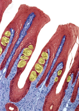
The figure above is an enhanced color light micrograph of a thin section through the tongue. The yellow colored structures are taste buds.
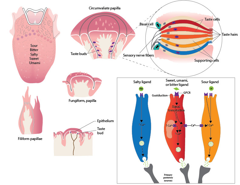
Four types of lingual papillae are observed on the human tongue: (1) circumvallate papillae (large groove surrounding the papilla), (2) fungiform papillae (mushroom shaped), (3) foliate papillae (leaf shaped), and (4) filiform papillae (string shaped) so named based on the shapes of each (see image below). Only the circumvallate, fungiform, and foliate papillae contain taste buds; the filiform provide a rough surface for moving food around in the mouth. On average, people have 2000 to 5000 taste buds, but in some exceptional cases, 20,000 have been observed. This has prompted some to propose taste classifications for people that include "non-tasters," "tasters," and "super tasters." Taste, however, is not an all or none phenomenon and the classifications of "non-taster" and "super tasters" simply represent populations of people on opposite ends of the spectrum. Interestingly, some "supertasters" taste and review food for a living and have insured their tasting abilities (job security) for astronomical amounts, like 1 million dollars.

© 2013 Encyclopædia Britannica, Inc. Downloaded from Image Quest Britannica; BYU-Idaho.
Moving from left to right, the first figure is an artificially colored scanning electron micrograph (SEM) of a tongue (rat). The second figure is an SEM of the tongue illustrating circumvallate papillae, the third figure is an SEM of fungiform papillae and the fourth figure is a light micrograph of filiform papillae.
Physiology of taste
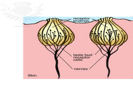
Each taste bud contains 50-150 receptor cells (taste or gustatory cells) sandwiched in between supporting cells and basal cells. Receptor cells are specialized epithelial cells that synapse with neurons. They have the ability to excite their associated neuron when they interact with a taste stimulus, generating action potentials in the neurons. The receptor cells have a relatively short life span and must be replaced about every 10 days. The role of the basal cells in the taste buds is to replace the worn out receptor cells. The taste stimulants (tastants) that react with receptor cells most efficiently are: sodium chloride (salty), sucrose (sweet), hydrochloric acid (sour), quinine (bitter) and monosodium glutamate (umami). There is a unique receptor for each of these taste modalities. However, other molecules can also interact with these receptors. For example, artificial sweeteners (e.g., saccharin and aspartame) are known to be 10,000 to 100,000 times more effective than sucrose at simulating the sweet receptors. Each receptor cell is oriented in the taste bud such that the tip of the cell protrudes into the oral cavity through an opening called a taste pore. This orientation allows the apical end to respond to the tastant and the basal end to transmit the signal to the brain.

© 2013 Encyclopædia Britannica, Inc. Downloaded from Image Quest Britannica; BYU-Idaho.
Illustration of a typical taste bud containing the taste cells.
As with all sensory systems, taste receptor cells must somehow convert an external or environmental stimulus into an internal or cellular response. In the body, these cellular responses are often associated with changes in membrane potentials. When the receptor is a non-neural cell, as is the case with taste, the depolarizations are called receptor potentials. The receptor potential is not an action potential but a graded potential that can modulate the activity of ion channels and trigger an action potential in a neuron. Typically the neurons that are associated with taste cells have a resting membrane potential of around -70mV. In order for a response (an action potential) to be generated something has to cause the membrane potential of the neuron to reach threshold. In the case of taste, a neurotransmitter is released from the basal membrane of the taste receptor cell. Release of the neurotransmitter is triggered when a tastant interacts with apical surface of the receptor cell. This interaction generates a depolarizing receptor potential that causes voltage-gated calcium channels on the basal surface of the cell to open. Calcium diffuses into the cell and acts as a signal to trigger exocytosis, releasing the neurotransmitter onto the neural cells. The neurotransmitter then elicits and action potential in the neuron. (Note: If you don't remember how a chemical synapse works review module 6.) Even though different taste receptor cells respond to different tastants, the release of a neurotransmitter from the receptor cells is always caused by an increase in intracellular Ca++. Thus, to understand how a chemical tastant (external stimulus) results in a neuronal signal (internal response) we need to understand how the tastant can cause a depolarization of the receptor cell and thus an increase in intracellular Ca++.
For a substance to be tasted, it must first dissolve in the saliva of the mouth. Once dissolved, the substance can interact with the apical membrane of a specific receptor cell. Receptor cells that respond to salt or sour substance contain membrane protein channels that allow the salty ion (Na+) or the sour ion (H+) to pass directly through the channel.

Image by Jared Cardinet BYU-Idaho F15
Signal transduction in a cell in response to a given taste stimulus.
This increased movement of positively charged ions across the membrane causes depolarization which in turn causes voltage sensitive Ca++ channels to open. Sensations of bitter, sweet and umami all use G-protein coupled receptors, thus, these substances don't cross the membrane, but the effect is still the same (Ca++increases in the cell which facilitates the release of neurotransmitter).
At least three different properties of the taste sensation pathway allow the brain to distinguish between different taste sensations: (1) the proportion of action potentials received from the different receptor types, (2) additional input from smell, and (3) additional input from other types of sensory receptors in the mouth. More specifically, foods that contain a higher proportion of salty to sweet chemicals will taste more salty. The sense of smell is very important to our perception of taste. Think of how food tastes when you have a cold and your nasal passageways are congested. Without the input of smell, many foods taste bland, for example, an onion tastes more like an apple. Other input from receptors that detect temperature, texture, and pain enhance the perception of the food. An example of this enhancement is the chemical capsaicin. This chemical is found in varying quantities in different kinds of peppers, endowing them with spiciness. However, our perception of spiciness is not the result of activation of a specific taste receptor but is due to the activation of pain receptors. Still, this enhanced taste sensation from capsaicin is a must for many people.
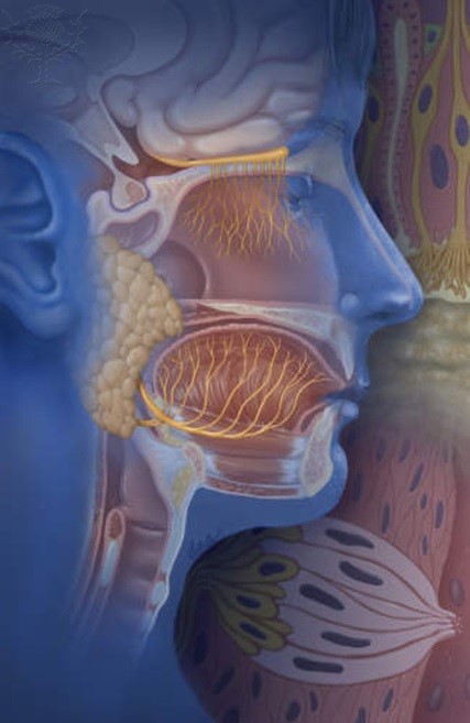
The perception of taste is produced by neural processing within the primary sensory cortex of the brain as it receives action potentials from different nerve bundles. The three major nerves that contribute to gustation are the facial nerve, which receives input from taste receptors located on the anterior two thirds of the tongue, the glossopharyngeal nerve, associated with the posterior one third of the tongue, and the vagus nerve, associated with the surface of the epiglottis. All of the nerves converge on the solitary nucleus of the medulla oblongata and eventually arrive to the lateral regions of the primary somatosensory cortex.

© 2013 Encyclopædia Britannica, Inc. Downloaded from Image Quest Britannica; BYU-Idaho.
Image shows the location of the facial and glossopharyngeal neurons innervating the tongue. The facial is the large neuron extending along the length of the jaw. The glossopharyngeal innervates the most posterior aspects of the tongue. This image also shows the olfactory neuron at the top. Olfaction is also very important for the experience of taste.
**You may use the buttons below to go to the next or previous reading in this Module**

