The Brain
THE BRAINSTEM AND CEREBELLUM
Brainstem
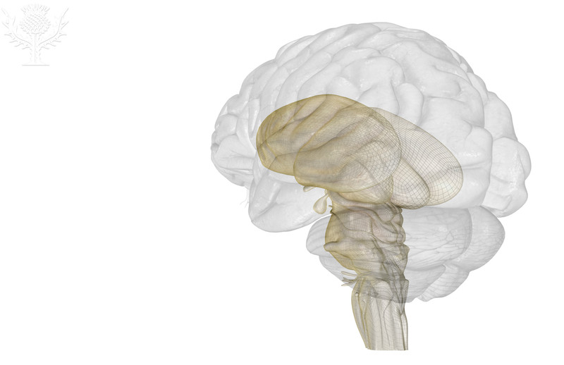
The region of the brain that connects the brain to the spinal cord is the brain stem. The brain stem is subdivided into three regions: the midbrain, the pons, and the medulla oblongata. The brain stem is also the site where groups of axons (nerve tracts) either exit the brain as cranial nerves or continue on into the spinal cord.Indeed, ten of the twelve pairs of cranial nerves (III - XII) exit the central nervous system from the brain stem. It should also be noted the brain stem is essential for maintain critical body functions, such as respiration and regulation of the heart, and that we cannot survive without its functions. We can survive without a cerebrum, although we would not be conscious that we were alive, but the body cannot survive without the brain stem.

© 2013 Encyclopædia Britannica, Inc. Downloaded from Image Quest Britannica; BYU-Idaho.

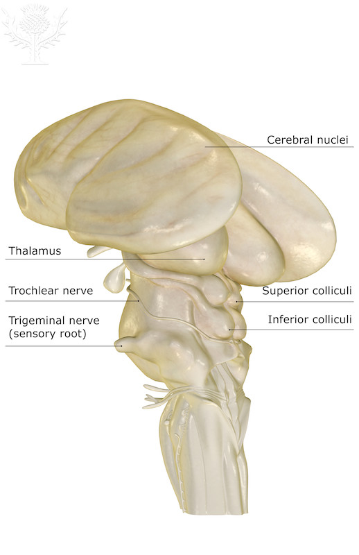
© 2013 Encyclopædia Britannica, Inc. Downloaded from Image Quest Britannica; BYU-Idaho.
The midbrain: The midbrain is the upper most portion of the brain stem and is situated between the diencephalon and the pons. Running through the midbrain is a hollow tube which connects the third and fourth ventricles (The ventricles of the brain are hollow spaces filled cerebral spinal fluid). Three unique clusters of cell bodies (nuclei) are observed in the midbrain; the corpora quadrigemina, the substantia nigra, and the red nucleus.
Spend time looking at the image search on the midbrain.
The copora quadrigemina are subdivided into two regions, the two superior colliculi and the two inferior colliculi. The paired superior colliculi (colluculus; singular) coordinate the movement of our eyes as we track a moving object. This reflex involves input from the eyes via the optic nerves, integration of the signal in the superior colliculi, and efferent signals to the muscles that control eye movements via cranial nerves III, IV and VI. The inferior colliculi help coordinate head and eye movements in respond to sudden sounds that cause you to abruptly move your head and turn your eyes toward the sound. For example, “Jaws,” or “Watcher in The Woods” kind of stuff.
The substantia nigra (dark substance) gets its name from its dark appearance in fresh tissue. This is due to the pigment neuromelanin (similar to melanin) which is produced in these cells. The neurons from this region produce dopamine as their neurotransmitter and neuromelanin is derived from the same precursor that produces melanin, L-dopa. Neurons from the substantia nigra ascend to the cerebrum and synapse with structures of the basal nuclei, a part of the brain involved in skeletal muscle control. Degeneration of these neurons is the cause of Parkinson’s disease, a condition in which the patient is unable to suppress unwanted muscle contractions resulting in constant tremors of the extremities.
Neurons in the red nucleus contain inordinate amounts of iron which when oxidized conveys a red hue. In many vertEbrates, it is a relay center for motor pathways that effect limb flexion. It is thought that in humans; the red nucleus influences arm swing during gait, crawling in babies and motor control of some of the larger shoulder and arm muscles, but not the legs.
The Pons: The pons region of the brain stem contains nuclei that contribute to the control of sleep, respiration, swallowing, bladder control, hearing, equilibrium, taste, eye movement, facial expressions, and posture. It may also play a role in generating dreams. In addition the pons (which means bridge) connects the cerebellum to the rest of the brain.
The Medulla Oblongata: "Alligators are ornery cause of their Medulla Oblongata"! (Water boy, 1998). Actually, the medulla oblongata probably has nothing to with ornery … surprise! As if movies ever tell the truth … Anyway, the medulla oblongata has a crucial role in body homeostasis. Neurons in the medulla oblongata adjust the force and rate of heart contractions, they generate and modify depth of breathing and regulate other fun activities like vomiting, hiccupping, coughing and sneezing. Additionally, the medulla is where most of the neurons that control voluntary skeletal muscle contraction cross over to the opposite side of the brain stem. This result of this crossing over is that the right side of the brain controls muscles on the left side of the body and the left side of the brain controls muscles on the right side of the body. The structure in the medulla where this crossing over is the olives of the medulla. The technical name for crossing over is decussation. This word comes from "deca" the prefix for the number 10 and the Roman Numeral for 10 which is X. The symbol X implies crossing over.
Cerebellum
The cerebellum ("little brain") sits under the occipital lobes of the cerebral hemispheres and attaches to the brain stem. Its structure is similar to the cerebrum in that it has a cortex composed of gray matter with white matter in the center. Likewise, the surface of the cerebellum is composed of folds called folia. It functions in motor learning, motor coordination and equilibrium. Smooth, coordinated skeletal muscle movement requires a functioning cerebellum. As we practice and learn complex movements the cerebellum is crucial in fine tuning the movements. When the motor cortex of the frontal lobes sends orders to the muscles to perform a particular task a copy of those orders is sent to the cerebellum. It, in turn, receives feedback from the proprioceptors in the muscles and joints as well as information from the inner ear relating to balance and equilibrium and compares what is actually happening with what the motor cortex ordered. Based on these comparisons, information is relayed back to the motor cortex in the cerebrum to fine tune the motor activity. The end result is the development of smooth, coordinated movements and agility for tasks like: typing, driving, piano playing, dancing etc. In addition, procedural memories for tasks like how to walk or ride a bike are stored here. Other studies using brain imaging and observations of patients with cerebellar injuries suggest that the cerebellum also plays roles in language, thought processing, and emotions.
**You may use the buttons below to go to the next or previous reading in this Module**


